|
 Previous Event | Next Event Previous Event | Next Event 
1955: DNA Copying Enzyme
Arthur Kornberg and his colleagues isolated DNA polymerase, an enzyme later used for all kinds of recombinant DNA techniques and for sequencing.
Kornberg’s group isolated DNA polymerase from E. coli bacteria. When he added the protein to a tube of salt solution containing DNA molecules and nucleotide building blocks, it was able to synthesize new strands of DNA.
The enzyme they had isolated was actually one of three forms of DNA polymerase in E. coli. The one Kornberg’s group found is chiefly used to repair DNA. His son Roger later found the polymerase that has the main job of replicating E. coli DNA when a cell divides.
In modern molecular biology labs, purified DNA polymerase is used routinely – to copy DNA by PCR (the polymerase chain reaction), for various recombinant DNA techniques, and to run sequencing reactions.
More Information
Reference:
A. Kornberg, "DNA Synthesis". San Francisco: Freeman, 1974.
Kornberg was awarded the 1959 Nobel Prize in Physiology or Medicine, along with Severo Ochoa, "for their discovery of the mechanisms in the biological synthesis of ribonucleic acid and deoxyribonucleic acid".
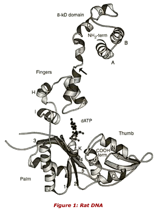
Structure of DNA POL:
FIG. 1 in Science June 24 1994 Sawaya et al. "Structure of Rat DNA Pol Beta". p 1930.
FIG. 1. Ribbon drawing (57) of rat DNA polymerase 3. The individual domains-subdomains are composed of the following residues: 8-lcD domain (1-87), fingers (88-151), palm (152-262), thumb (263-335). The arrow marks the location of the proteolytically sensitive hinge between the 31-kD catalytic domain and the 8-kD template binding domain. The ribbon diagram KF and HIV RT are shown in the same orientation in (14). T7 RNAP is shown in (16). All four polymerases binding channels composed of fingers, palm, and thumb regions. The 8-kD domain has no counterpart in KF, RT, or RNAP.
Previous Event | Next Event
Last Reviewed: April 14, 2008
|