
"Imaging technology is used across the entire spectrum, from very basic research to clinical care," says Dr. James Tatum, associate director of NCI's Cancer Imaging Program (CIP), which is part of the Division of Cancer Treatment and Diagnosis and currently funds a portfolio of over 400 grant projects.
Part of CIP's mandate has been to support areas of cancer-imaging research considered too high-risk for commercial investment. Through its Imaging Development Group, it helps shepherd promising new imaging compounds from discovery through early clinical testing. Several of these new agents are now in clinical trials across the
country, with more in the pipeline.
To speed up and improve early clinical trials of these and other promising new imaging agents, CIP has been working closely with the American College of Radiology Imaging Network (ACRIN), an NCI-funded cooperative group.
Best known for performing large trials of mature imaging technology, such as the National CT Colonography Trial, ACRIN has received assistance from CIP to conduct early multicenter trials as well, explains Dr. David Mankoff, professor of radiology at the University of Washington and an ACRIN investigator. Examples of agents being tested in these smaller trials, coordinated through ACRIN's Experimental Imaging Sciences Committee, include 64Cu-ATSM to measure treatment response in cervical cancer, and 18F-fluoromisonidazole to monitor brain-tumor hypoxia (decreased oxygen supply).
A huge challenge in trials of these and other imaging radioisotopes is that "many of these probes have extremely short half lives, about two hours or so," explained Dr. Mankoff. "You can't just make them in one place and ship them all over the country - you have to have a distribution network." Fortunately, the clinical success of FDG-PET imaging has led to a commercial distribution network to make and distribute the fragile FDG isotope to clinical sites across the country. CIP "has been key" in convincing members of this network to also manufacture experimental probes for ACRIN, said Dr. Mankoff.
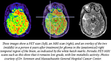
CIP also helps fund advanced imaging research laboratories around the country, including one that has the first dual MRI-PET device in the country, commissioned by Dr. Gregory Sorensen and colleagues at Massachusetts General Hospital.
"We are now able to combine the high spatial resolution and functional information from MRI with the metabolic and receptor information available from PET, to more carefully study tumor hypoxia, angiogenesis, and the link between [tumor] metabolism and response to therapies," explained Dr. Sorensen, who will soon begin the first clinical trial (funded in part by CIP) of dual MRI-PET to monitor patients with brain tumors during treatment.
Imaging technology is also poised to help researchers visualize the intricate inner workings of cancer cells. To this end, CIP has funded eight In vivo Cancer Molecular Imaging Centers that will help advance cellular and molecular imaging related to cancer.
"There is a compelling need to understand cancer from a systems biology perspective, and tumor biology is the most complex system you could possibly imagine," says Dr. Tatum. "It is my own belief that the only way you can understand what goes on in these complex systems in vivo is to be able to monitor or interrogate the system non-invasively employing advanced imaging methods across the resolution from cells to patients."
| 