
The Visible Human Project
Getting the Data
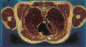
The initial aim of the Visible Human Project® is to create a digital image dataset of complete human male and female cadavers in MRI, CT and anatomical modes.The male dataset consists of axial MR images of the head and neck taken at 4 mm intervals and longitudinal sections of the remainder of the body also at 4 mm intervals. The resolution of the MR images is 256 pixels by 256 pixels. Each pixel has 12 bits of grey tone.
The CT data consists of axial CT scans of the entire body taken at 1 mm intervals at a resolution of 512 pixels by 512 pixels where each pixel is made up of 12 bits of grey tone. The axial anatomical images are 2048 pixels by 1216 pixels where each pixel is defined by 24 bits of color, each image consisting of about 7.5 megabytes of data. The anatomical cross-sections are also at 1 mm intervals and coincide with the CT axial images. There are 1871 cross-sections for each mode, CT and anatomy, obtained from the male cadaver.
The dataset from the female cadaver has the same characteristics as the male cadaver with one exception. The axial anatomical images were obtained at 0.33 mm intervals instead of 1.0 mm intervals. This results in over 5,000 anatomical images. The female dataset is about 40 gigabytes in size. Spacing in the "Z" direction was reduced to 0.33 mm in order to match the pixel spacing in the "XY" plane which is 0.33 mm. This enables developers who are interested in three-dimensional reconstructions to work with cubic voxels.
A recent addition to the male dataset is the inclusion of higher resolution anatomical images. 70mm film taken during the original data collection phase has been digitized at a resolution of 4096 pixels by 2700 pixels where each pixel is made up of 24 bits of color. As with the original anatomical images, there are a total of 1871 of these high resolution images.
A single License Agreement covering use of both the male and female Visible Human Project® datasets is available, as a text file, Word file, or a PDF file. Please make two copies of the agreement and have both signed as originals by your appropriate officials. The agreement requires that you include a brief statement explaining your intended use of the dataset. Please prepare this statement and include your return postal and email address. This statement must be included for the agreement to be processed. Send both signed copies of the agreement and the statement of how you intend to use the data to:
- Visible Human Project®
National Library of Medicine
Building 38A, Room B1N-30
8600 Rockville Pike
Bethesda, MD 20894
The agreement will be signed by the NLM and one of the originals will be returned to you. Included with the signed agreement will be your account and password to the Visible Human Project® FTP site. On the FTP site are folders containing the following:
- Radiological: The original radiological images, in a lossless "Z" compression, in the GE format as received from the MRI and CT scanners, text files of the associated MRI and CT headers, and instructions on how to open these files.
- Full Color: The original cryosectional images at 0.33mm XY resolution, in a lossless "Z" compression, in a .raw format, and instructions on how to open these files.
- 70mm: The cryosectional images at 0.17 XY resolution derived from scanning the original 70mm film, in a lossless "Z" compression, in a .rgb format, and instructions on how to open these files.
- PNG_format: The original radiological images and fullcolor images formatted in a lossless, directly readable .png format, and text files of the associated MRI and CT headers.
We recommend that you use the .png files. Each file contains an individual image in PNG (Portable Network Graphics) format. The anatomical images will appear directly when opened by your viewer. The radiological images (CT, MRI and X-ray) will appear as a black screen. These images are 16 bit grayscale. Because there is actually only up to 10 bits of data in each pixel, the images will appear to be black when initially loaded into your viewer. To properly view the images, use the "auto adjust" feature of your viewer or manually adjust the gamma level. The CT, MRI or X-ray image will now be visible.
The male and female data sets are also available to NLM licensees on DVD from the National Technical Information Service (N.T.I.S.) at http://www.ntis.gov/products/vishuman.aspx. An order form will be included with your signed copy of the Agreement.
Sample full scale images from the Male and Female Datasets are available via the NLM FTP site, including:
- Eleven full color anatomical images in .png format: 6 from the Visible Male, and 5 from the Visible Female. Please note that each of these image downloads is approximatly 3.5 megabytes in size, the full opened image being about 7 megabytes in size.
- Ten CT scan images in .png format: 5 images captured before the cadaver was frozen, and 5 images captured after it was frozen.
- Eighteen MRI images in .png format: six T1 images, six T2 images and six proton density images.
Your continued interest in the Visible Human Project® is greatly appreciated. Comments cncerning any aspect of the data set would be welcome. Please e-mail your suggestions to:
- Michael J. Ackerman, Ph.D.
Project Officer
vhp@nlm.nih.gov