The notion that some substances in the environment can
damage the nervous system has an ancient history. The neurotoxicity
of lead was recognized more than 2,000 years ago by the
Greek physician Dioscerides, who wrote, “Lead makes
the mind give way.” In the intervening millennia
many other substances have been added to the list of known
or suspected neurotoxicants. Despite this accumulation
of knowledge, there is still much that isn’t understood
about how neurotoxicants affect the developing brain, especially
the effects of low-dose exposures. Today researchers are
taking a hard look at low-dose exposures in utero and
during childhood to unravel some of the mysteries of impaired
neurodevelopment.
About 17% of school-age children in the United States
suffer from a disability that affects their
behavior, memory, or ability to learn, according to a study
published in
the March 1994 issue of Pediatrics by a team from
the Centers for Disease Control and Prevention
(CDC). The list of maladies includes attention deficit/hyperactivity
disorder (ADHD), autistic spectrum disorders,
epilepsy,
Tourette syndrome, and less specific conditions
such as mental retardation and cerebral palsy. All
are believed to be the outcome of some abnormal process
that unfolded
as the brain was developing in utero or in
the young child.
 |
| image: Ahmed Hussam/iStockphoto |
These disorders have an enormous impact on families and
society. According to the 1996 book Learning Disabilities:
Lifelong Issues, children with these disorders have
higher rates of mental illness and suicide, and are more
likely to engage in substance abuse and to commit crimes
as adults. The overall economic cost of neurodevelopmental
disorders in the United States is estimated to be $81.5-167
billion per year, according to a report published in the
December 2001 issue of EHP Supplements.
Potentially even more disturbing is that a number of
epidemiologic studies suggest that the incidence of certain
disorders is on the rise. In the United States, the diagnosis
of autistic spectrum disorders increased from 4-5 per 10,000
children in the 1980s to 30-60 per 10,000 children in the
1990s, according to a report in the August 2003 Journal
of Autism and Developmental Disorders. Similarly, notes
a report in the February 2002 issue of CNS Drugs,
the diagnosis of ADHD grew 250% between 1990 and 1998.
The number of children in special education programs classified
with learning disabilities increased 191% between 1977
and 1994, according to an article in Advances in Learning
and Behavioral Disabilities, Volume 12, published in
1998.
So what is going on? The short answer is that no one
really knows. There’s not even consensus on what
the soaring rates actually mean. Heightened public awareness
could account for the surge in the numbers, or it may be
that physicians are getting better at diagnosing the conditions.
Some autism researchers believe the rise in that condition’s
prevalence simply reflects changes in diagnostic criteria
over the last 25 years. On the other hand, some scientists
believe that the rates of neurodevelopmental disease are
truly increasing, and that the growing burden of chemicals
in the environment may play a role.
With that in mind, investigators are considering the
effects of gene-environment interactions. A child with
a mild genetic tendency toward a neurodevelopmental disorder
might develop without clinically measurable abnormalities
in the absence of environmental “hits.” However,
children in industrialized nations develop and grow up
in a veritable sea of xenobiotic chemicals, says Isaac
Pessah, director of the University of California, Davis,
Center for Children’s Environmental Health and Disease
Prevention. “Fortunately,” he says, “most
of us have a host of defense mechanisms that protect us
from adverse outcomes. However, genetic polymorphisms,
complex epistasis, and cytogenetic abnormalities could
weaken these defenses and amplify chemical damage, initiating
a freefall into a clinical syndrome.”
 |
| image: Photodisc |
Pessah cites the example of autism. He says susceptibility
for autism is likely conferred by several defective genes,
no one of which can account for all the core symptoms of
social disinterest, repetitive and overly focused behaviors,
and problems in communication. Could multiple genetic liabilities
and exposure to a chemically complex environment act in
concert to increase the incidence and severity of the condition?
Despite the uncertainties, many scientists believe it
would be wise to err on the side of caution when it comes
to a research agenda. As Martha Herbert, a pediatric neurologist
at Harvard Medical School, puts it, “Even though
we may have neither consensus nor certainty about an autism
epidemic, there are enough studies coming in with higher
numbers that we should take it seriously. Environmental
hypotheses ought to be central to research now. The physiological
systems that have been harmed by environmental factors
may also point to treatment targets, and this might be
a great way to help the children.”
The Parade of Neurotoxicants
Among the most intensely studied neurotoxicants are metals
(lead, mercury, and manganese), pesticides, polychlorinated
biphenyls (PCBs), and polybrominated diphenyl ethers (PBDEs).
A number of these compounds were identified as neurotoxicants
when individuals were exposed to high doses during occupational
accidents or childhood poisonings. Scientists are now exploring
the potential consequences of low-dose exposures, especially
to children and fetuses. Epidemiologic studies play a central
role, and these are often complemented by experimental
work on animals and cell cultures. These days, researchers
are looking not only at associations between toxicants
and disease, but also at the underlying cellular and molecular
mechanisms.
Lead. Studies dating to the 1970s show
that children exposed to lead have deficits in IQ, attention,
and language. In response, the CDC revised its limits for
acceptable blood levels of the metal in several steps,
from 60 micrograms per deciliter (µg/dL) in the 1960s
to the current level of 10 µg/dL, set in 1991. But
many scientists think that limit is still too high. A study
reported in the September 2005 issue of EHP found
that there were significant effects on a child’s
IQ even when blood lead concentrations were below 10 µg/dL.
Upon the July 2005 release of the Third National Report
on Human Exposure to Environmental Chemicals by
the CDC, Jim Pirkle, deputy director for science at the
CDC’s Environmental Health Laboratory, stated, “There
is no safe blood [lead] level in children.”
Several groups have also found evidence that lead exposure
may shape a child’s social behavior. An article in
the May 2000 issue of Environmental Research reports
a strong correlation, dating back to 1900, between violent
crime and the use of lead-based paint and leaded gasoline.
The research complements studies by Herbert Needleman,
a professor of psychiatry and pediatrics at the University
of Pittsburgh School of Medicine, who found that bone lead
levels in young males were correlated with aggression and
criminality. “Lead is significantly associated with
a risk for delinquency,” says Needleman. His research
appeared in the November-December 2002 issue of Neurotoxicology
and Teratology and the 7 February 1996 issue of JAMA.
Another new area of research links early lead exposure
to changes in the aging brain. Nasser Zawia, an associate
professor of pharmacology and toxicology at the University
of Rhode Island, Kingston, and his colleagues found increased
expression of amyloid precursor protein (APP) and its product, β-amyloid
(which is a hallmark of Alzheimer disease), in aging rats
that were exposed to lead shortly after birth. In contrast,
old rats that were exposed to lead did not show an increased
expression of APP and β-amyloid.
The work, published in the 26 January 2005 issue of The
Journal of Neuroscience, suggests that early exposure
to lead can “reprogram” gene expression and
regulation later in life. According to Zawia, preliminary
research also shows that “monkeys exposed to lead
as infants exhibit similar molecular changes as well as
exaggerated Alzheimer’s pathology.”
 |
| image: Photodisc |
Mercury. The current Environmental Protection
Agency (EPA) reference dose for methylmercury (an organic,
toxic form of mercury) is 0.1 micrograms per kilogram per
day (µg/kg/day). Humans are exposed to methylmercury
primarily through consumption of contaminated fish; a good
70% of this contamination comes from anthropogenic sources
such as emissions from coal-fired power plants. High-level
exposure to methylmercury in the womb is linked to a number
of impairments, including mental retardation, cerebral
palsy, seizures, deafness, blindness, and speech difficulties.
An article in the May 2005 issue of EHP puts
the economic cost to the United States of methylmercury-induced
toxicity (in terms of lost productivity) at $8.7 billion
annually.
The effects of low-dose exposures are not so apparent.
Two large epidemiologic studies of fishing populations
in the Faroe Islands and the Seychelles have produced conflicting
results regarding low-dose effects. Both studies sought
to examine the association between methylmercury exposure
and neurodevelopment in children whose mothers ate contaminated
seafood during pregnancy.
The leader of the Faroe Islands study, Philippe Grandjean,
an adjunct professor of environmental health at the Harvard
School of Public Health, and his colleagues reported in
the November 1997 issue of Neurotoxicology and Teratology that
7-year-old Faroese children had significant cognitive deficits
and neurological changes after prenatal exposure to methylmercury.
Grandjean’s team followed up on the children at age
14. According to a report in the February 2004 issue of The
Journal of Pediatrics, the children continued to have
problems, including neurological changes and decreased
nervous control of the heart.
In contrast, the authors of the Seychelles study found
little evidence of lasting harm on a cohort
of 66-month-old children, according to their report in
the 26 August 1998
issue of JAMA. A follow-up study, published in the
17 May 2003 issue of The Lancet, similarly
found no lasting effects on language, memory,
motor skills, or behavioral function when the children
were 9 years old.
The different outcomes of the two studies are puzzling
because the children of both populations appeared to be
exposed to similar amounts of methylmercury. Several explanations
have been proposed, including the possibility that genetic
differences between the populations may alter their relative
predispositions to harm from mercury exposure. The source
of methylmercury is also different in the two populations.
The Faroese are exposed primarily through the consumption
of pilot whale meat, whereas the Seychelles population
relies heavily on ocean fish. According to Gary Myers,
a professor of neurology and pediatrics at the University
of Rochester Medical Center and one of the principal investigators
of the Seychelles study, whale meat contains many other
contaminants (including PCBs) besides methylmercury. “There
is also evidence,” he says, “that the effects
of concomitant PCB and mercury exposure are synergistic.”
Researchers continue to look at whether there is a danger
from methylmercury at the levels of exposure achieved by
fish consumption. Another layer of uncertainty was added
with findings published in the October 2005 issue of EHP showing
that fish consumption during pregnancy appeared to boost
infant cognition--but only as long as mercury intake, as
measured in maternal hair, wasn’t too high.
 |
| image: RMAX/iStockphoto |
The question of whether low levels of mercury are harmful
has also manifested itself in a controversy over the use
of vaccines containing thimerosal, a preservative. Although
thimerosal was removed from many of these vaccines in 2001,
children that were immunized before that date could have
received a cumulative dose of more than 200 µg/kg
of mercury with the routine complement of childhood vaccinations,
according to a study in the May 2001 issue of Pediatrics.
Thimerosal is nearly half ethylmercury by weight. Because
ethylmercury is an organic form of mercury, there is some
suspicion that it acts like methylmercury in the brain,
although research published in the August 2005 issue of EHP suggests
that the two forms differ greatly in how they are distributed
through and eliminated from the brain. Developing countries
continue to use pediatric vaccines that contain thimerosal.
In the United States, thimerosal is still present in influenza
vaccines, which the CDC recommends be given to pregnant
women and children aged 6-23 months.
Advocacy groups, such as SafeMinds, have suggested that
the decades-long rise in the diagnosis of autism is related
to the presence of thimerosal in vaccines. In May 2004,
however, the Institute of Medicine (IOM) issued a report, Immunization
Safety Review: Vaccines and Autism, stating that several
epidemiological studies published since 2001 “consistently
provided evidence of no association” between thimerosal-containing
vaccines and autism. However, the IOM’s report has
been severely criticized by a number of advocacy groups,
including the National Autism Association, for relying
too heavily on a specific set of epidemiologic data while
dismissing clinical evidence and other epidemiologic studies
that showed evidence of a link.
Despite the assurances of the IOM, some scientists continue
to explore the mechanisms underlying the potential neurotoxic
effects of thimerosal. In the January 2005 issue of NeuroToxicology,
S. Jill James, a professor of pediatrics at the University
of Arkansas for Medical Sciences, and her colleagues report
that the neuronal and glial cell toxicity of methylmercury
and ethylmercury (as dosed via thimerosal) are both mediated
by the depletion of the antioxidant peptide glutathione.
Of the two cell types, neurons were found to be particularly
susceptible to ethylmercury-induced glutathione depletion
and cell death, according to James, and pretreatment of
the cells with glutathione reduced these effects. Other
studies by James and her colleagues, reported in the December
2004 issue of the American Journal of Clinical Nutrition,
showed that autistic children had lower levels of glutathione
compared to normal controls, and may therefore have had
a significant reduction in the ability to detoxify reactive
oxygen species.
James says the abnormal profile “suggests that
these children may have an increased vulnerability to pro-oxidant
environmental exposures and a lower threshold for oxidative
neurotoxicity and immunotoxicity.” Speaking at the
XXII International Neurotoxicology Conference in September
2005, she presented evidence that multiple genetic polymorphisms
affecting glutathione pathways may interact to produce
a chronic metabolic imbalance that could contribute to
the development and clinical symptoms of autism. Her paper
in the American Journal of Clinical Nutrition reported
that low glutathione levels in many autistic children were
reversible with targeted nutritional intervention, but
the ramifications of this finding are still unclear.
 |
| image: Shutterstock |
Manganese. As an essential nutrient, manganese
is required for normal development; the reference dose
for manganese is 0.14 mg/kg/day. Chronic occupational exposure
to high levels of this metal is associated with manganism,
a condition reminiscent of Parkinson disease that is characterized
by tremors, rigidity, and psychosis. The illness is seem
primarily among miners.
Animal studies published in the August 2005 issue of Neurotoxicology by
David Dorman, director of the division of biological sciences
at the CIIT Centers for Health Research in Research Triangle
Park, North Carolina, suggest that the fetus is protected
to a certain extent from maternally inhaled manganese.
According to Dorman, children are exposed to manganese
primarily by ingesting it, but he knows of no link between
childhood exposure to manganese and later Parkinson disease.
Nevertheless, because manganese affects the adult brain,
people suspect that the developing brain may be even more
susceptible to harm from this metal, and recent research
has unveiled a new cause for concern: In the January 2006
issue of EHP, child psychiatry professor Gail Wasserman
and colleagues from Columbia University reported that Bangladeshi
children who drank well water with high concentrations
of naturally occurring manganese had diminished intellectual
function. The researchers noted that the bioavailability
of manganese in water is higher than that of manganese
in food. They also pointed out that about 6% of U.S. wells
have a high enough manganese content to potentially put
some children at risk for diminished intellectual function.
The cellular and molecular mechanisms of manganese neurotoxicity
are not well understood. The dopaminergic system in the
basal ganglia, which is affected in Parkinson disease,
may be involved, but this hypothesis is controversial.
Tomás Guilarte, a professor of molecular neurotoxicology
at the Johns Hopkins Bloomberg School of Public Health,
described research on these systems in nonhuman primates
at the XXII International Neurotoxicology Conference. According
to Guilarte, unpublished positron-emission tomography studies
of the basal ganglia show that “manganese does appear
to have an effect on dopaminergic neurons.” Guilarte
found that the more manganese the animals received, the
less dopamine was released through the actions of amphetamine
(which is used to induce the release of the neurotransmitter). “This
does not mean that manganese causes Parkinson’s disease,
merely that it has an effect on those neurons,” he
says. This is the first report of an in vivo effect
on dopamine release by manganese.
PCBs, PBDEs, and pesticides. Many chemicals
raise concerns because of their persistence in the environment
and their tendency to bioaccumulate in animal tissues.
They are typically synthetic molecules that were designed
for use in everyday products, such as electrical equipment,
computers, furniture, and pesticides.
PCBs appear to be present in all parts of the food chain,
and humans are exposed to these molecules primarily through
the ingestion of animal fat. The toxicity of these chemicals
was first recognized after mass poisonings in Japan in
1968 and Taiwan in 1979. Children born to women who had
ingested contaminated cooking oil in Taiwan had a number
of developmental abnormalities, including psychomotor delay
and lower scores on cognitive tests, according to a report
in the 15 July 1988 issue of Science.
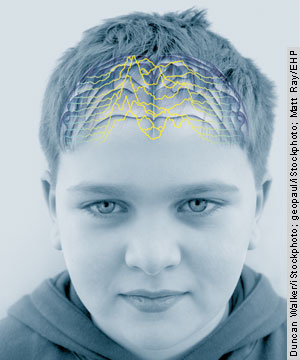
 |
| image: Duncan Walker/iStockphoto |
Since those earlier observations, several studies have
described a connection between prenatal exposure to PCBs
and delayed cognitive development and lower IQ. For example,
a study in the 10 November 2001 Lancet reports those
infants and young children exposed to PCBs through breast
milk scored lower on tests of psychomotor and mental development.
The mothers were exposed to normal background levels of
PCBs in Europe. In response to such studies, the U.S. Food
and Drug Administration set tolerance levels for PCBs in
a number of consumer products, such as milk and manufactured
dairy products (1.5 parts per million), poultry (3.0 parts
per million), and baby food (0.2 part per million).
PBDEs are widely used as flame retardants in consumer
products. The effects of PBDEs on humans is not clear,
but animal toxicity studies described in volume 183 (2004)
of Reviews of Environmental Contaminants and Toxicology show
that PBDEs can cause permanent learning and memory impairments,
hearing deficits, and behavioral changes. There is a growing
concern about PBDEs because they appear to be accumulating
in human tissues. Andreas Sjödin, a toxicologist at
the CDC, and colleagues found a trend toward increasing
concentrations of PBDEs in human serum taken from sample
populations in the southeastern United States from 1985
through 2002, and in Seattle, Washington, from 1999 through
2002. This report appears in the May 2004 EHP. Several
studies have also discovered PBDEs in human breast milk.
The current EPA reference dose for PBDEs is 2 mg/kg/day.
As for pesticides, it’s been suggested by zoologist
Theo Colborn of the University of Florida that every child
conceived today in the Northern Hemisphere is exposed to
these chemicals from conception through gestation and beyond.
Some pesticides appear to be more harmful than others,
and so the reference dose varies somewhat from one compound
to another.
 |
| image: Corbis |
The effects of pesticides on the developing brain have
been investigated in human epidemiologic studies and in
laboratory experiments with animals. Vincent Garry, a professor
of environmental medicine at the University of Minnesota,
and his colleagues found that children born to applicators
of the fumigant phosphine were more likely to display adverse
neurological and neurobehavioral developmental effects.
The herbicide glyphosate was also linked to neurobehavioral
effects, according to the same report, which appeared in
the June 2002 issue of EHP Supplements. Another
epidemiologic study, reported in the March 2005 issue of NeuroToxicology, showed
that women who were exposed to organophosphate pesticides
in an agricultural community in California had children
who displayed adverse neurodevelopmental effects, and that
higher levels of pesticide metabolites in maternal urine
were associated with abnormal reflexes in the women’s
newborn children.
Many PCBs, PBDEs, and pesticides are the subject of the
2001 Stockholm Convention on Persistent Organic Pollutants,
which became international law in May 2004. The goal of
the treaty is to “rid the world of PCBs, dioxins
and furans, and nine highly dangerous pesticides,” according
to the United Nations Environment Programme. Implementation
of the treaty has significant practical challenges, however,
including the difficulty of eliminating one persistent
pollutant without creating another (for example, when burning
PCBs yields by-products such as dioxins and furans).
Not Immune to Harm
Exposure to a neurotoxicant may not be the only way to
disrupt the natural growth of the brain. Scientists are
now looking at the subtle physiological effects of immunotoxicants
and infectious agents on biological events during development.
It turns out that mothers who experience an infection
during pregnancy are at a greater risk of having a child
with a neurodevelopmental disorder such as autism or schizophrenia.
For example, prenatal exposure to the rubella virus is
associated with neuromotor and behavioral abnormalities
in childhood and an increased risk of schizophrenia spectrum
disorders in adulthood, according to an article in the
March 2001 issue of Biological Psychiatry. Rubella
has also been linked to autism: some 8-13% of children
born during the 1964 rubella pandemic developed the disorder,
according to a report in the March 1967 Journal of Pediatrics.
The same study also noted a connection between the rubella
virus and mental retardation.
 |
| image: Shutterstock |
Some epidemiologic studies have found an increased risk
of schizophrenia among the children of women who were exposed
to the influenza virus during the second trimester of pregnancy,
according to a report in the February 2002 Current Opinion
in Neurobiology. In the August 2004 Archives of
General Psychiatry, Ezra Susser, head of epidemiology
at Columbia University’s Mailman School of Public
Health, and his colleagues reported that the risk of the
mental disorder was increased sevenfold if the schizophrenic
patient’s mother had influenza during her first trimester
of pregnancy. A prospective birth cohort study in the April
2001 Schizophrenia Bulletin found that second
trimester exposure to the diphtheria bacterium also significantly
increased the risk of schizophrenia.
How might infectious agents cause these disorders? According
to John Gilmore, a professor of psychiatry at the University
of North Carolina at Chapel Hill, maternal infections during
pregnancy can alter the development of fetal neurons in
the cerebral cortex of rats. The mechanism is far from
clear, but signaling molecules in the mother’s immune
system, called cytokines, have been implicated. Speaking
at the XXII International Neurotoxicology Conference, Gilmore
described in vitro experiments showing that elevated
levels of certain cytokines--interleukin-1β,
interleukin-6 and tumor necrosis factor-alpha (TNF- )--reduce
the survival of cortical neurons and decrease the complexity
of neuronal dendrites in the cerebral cortex. “I
believe that the weight of the data to date indicates [that
the maternal immune response] can have harmful effects,” says
Gilmore.
)--reduce
the survival of cortical neurons and decrease the complexity
of neuronal dendrites in the cerebral cortex. “I
believe that the weight of the data to date indicates [that
the maternal immune response] can have harmful effects,” says
Gilmore.
Inflammatory responses in the mother may not be the only
route to modifying the fetal brain. The University of California,
Davis, Center for Children’s Environmental Health
and Disease Prevention is conducting a large study of autistic
children in California called CHARGE (Childhood Autism
Risks from Genetics and the Environment), which suggests
that the child’s immune system may also be involved.
According to Pessah, the study principal investigator,
children with autism appear to have a unique immune system. “Autistic
children have a significant reduction in plasma immunoglobulins
and a skewed profile of plasma cytokines compared to other
children,” he says. “We think that an immune
system dysfunction may be one of the etiological cores
of autism.”
He continues, “We know that many of the things
that kids are exposed to these days are immunotoxicants.
. . . We have evidence that ethylmercury and thimerosal
alter the signaling properties of antigen-presenting cells,
known as dendritic cells, at nanomolar levels.” Since
each dendritic cell can activate 250 T cells, any dysregulation
will be magnified, he says. “Add to that a genetic
abnormality in processing immune information, and there
could be a problem.”
 |
| image: Photodisc |
Such problems might extend to the central nervous system.
The brains of individuals who have a neurodevelopmental
disorder also show evidence of inflammation. In the January
2005 issue of the Annals of Neurology, Carlos Pardo,
an assistant professor of neurology and pathology
at the Johns Hopkins University School of Medicine, and
his colleagues
report finding high levels of inflammatory
cytokines (interleukin-6, interleukin-8, and interferon- )
in the cerebrospinal fluid of autistic patients.
Glial
cells, which serve as the brain’s innate immune system,
are the primary sources of cytokines in the central nervous
system. So it may not be surprising that Pardo’s
team also discovered that glia are activated--showing both
morphological and physiological changes--in postmortem
brains of autistic patients.
)
in the cerebrospinal fluid of autistic patients.
Glial
cells, which serve as the brain’s innate immune system,
are the primary sources of cytokines in the central nervous
system. So it may not be surprising that Pardo’s
team also discovered that glia are activated--showing both
morphological and physiological changes--in postmortem
brains of autistic patients.
The recognition that the immune system is involved in
neurodevelopmental disorders is changing people’s
perceptions of these conditions. “Historically, scientists
have focused on the role of neurons in all kinds of neurological
diseases,” Pardo says, “but they have generally
been ignoring the [glia].” He adds, “In autism,
it could be that the [glia] are responding to some external
insult, such as an infection, an intrauterine injury, or
a neurotoxicant.”
According to Pardo, it’s still not clear whether
the neuroimmune responses associated with autism contribute
to the dysfunction of the brain or whether they are secondary
reactions to some neural abnormality. “John Gilmore’s
work [showing that cytokines can be harmful to brain cells]
is quite interesting and important,” he says. “However, in
vitro studies may produce results that don’t
reflect what occurs under in vivo conditions. Cytokines
like TNF- may
be beneficial for some neurobiological functions at low
concentrations, but may be extremely neurotoxic at high
concentrations.”
Lending Brain Power to Exposure Assessment
The medical and scientific communities recognize the
colossal challenges involved in identifying the ultimate
causes of neurodevelopmental disorders. This is complicated
by the sheer numbers of potential exposures involved. More
than 67% of the nearly 3,000 chemical compounds produced
or imported in amounts exceeding 1 million pounds per year
have not been examined with even basic tests for neurotoxicity,
according to Toxic Ignorance, a 1997 analysis by
Environmental Defense.
In the past few years, several large projects have been
proposed, and funding by the NIH has been increased. For
example, the NIH boosted its support for autism research
from $22 million in 1997 to $100 million in 2004. In 2001,
the NIEHS and the EPA jointly announced the creation of
four new children’s environmental health research
centers (including the one at the University of California,
Davis), which focus primarily on neurodevelopmental disorders.
More recently, the proposed multibillion-dollar National
Children’s Study, which is cosponsored by the Department
of Health and Human Services and the EPA, has been designed
to follow nearly 100,000 children over the course of 21
years. The investigators plan to study the effects of environmental
factors on children’s growth and development, including
impacts on learning, behavior, and mental health. Study
investigators hope to enroll the first participants in
early 2007.
Scientists also see the need for designing better studies.
In neurodevelopmental studies, as in any other field, the
quality of a study is only as good as all of its parts.
Jean Harry, head of the NIEHS Neurotoxicology Group, says, “You
can have a valid assessment of behavior, but in the absence
of good exposure data, a causative association with environmental
factors will be compromised.”
 |
| image: Shutterstock |
In a bid to address the difficulties faced by epidemiologic
studies that look for neurodevelopmental effects from in
utero chemical exposure, a working group of 20
experts gathered in September 2005 under the auspices of
the Penn State Hershey Medical Center, coincident with
the XXII International Neurotoxicology Conference. The
goal of their day-long session was to develop a scheme
of best practices for the design, conduct, and interpretation
of future investigations, as well as the practical inclusion
of new technologies, such as imaging.
At one point in the dialogue, the group recognized that
perhaps the greatest challenge in these studies was determining
how to evaluate in utero exposures to environmental
chemicals. “Quite often the very nature of epidemiological
studies limits the ability to perform accurate exposure
assessments,” says Harry, who was part of the expert
group. “Such exposures may have occurred in the distant
past, they may have been unknown, or they may have been
in conjunction with many other compounds.”
The group therefore recommended that actual measurements,
even if indirect, are better than methods based on subject
recall. It also recommended that a well-defined hypothesis
should form the foundation of in utero studies for
assessing neurodevelopmental outcomes. “[These and
other] conclusions will move the science forward by describing
methods that should improve interstudy comparisons, and
they offer ways in which research results should be reported
to the scientific and medical communities,” says
Judy LaKind, an adjunct associate professor of pediatrics
at the Hershey Medical Center and a member of the workshop
steering committee. The complete workshop report will be
published in an upcoming issue of NeuroToxicology.
Imagining the Big Picture
The challenges of addressing neurodevelopmental disorders
are more than scientific. The difficulties come together
at a crossroads where the communication of knowledge, the
treatment of patients, and the regulation of potentially
toxic chemicals meet. Says Herbert, “Evidence-based
medicine has not yet developed standards for assessing,
or practices for treating, the impacts of chronic, multiple
low-dose exposures.” Rather than waiting, she says,
patients and parents of patients are turning to alternative
medicine to address their concerns.
That’s not always a good thing, especially when
patients and parents may be misinformed. Kathy Lawson,
director of the Healthy Children Project at the Learning
Disabilities Association of America, says there is a disconnect
between scientific knowledge and the public’s awareness
of ways to reduce the incidence of some disorders. “In
my visits to various organizations, I’ve discovered
that people are completely unaware that there is a connection
between environmental toxicants and their health,” she
says. “Even pediatricians often don’t know
about these things,” she adds.
Educating the public is only part of the solution. Elise
Miller, executive director of the nonprofit Institute for
Children’s Environmental Health, thinks that federal
regulatory agencies do not adequately protect children’s
health. “The Toxic Substances Control Act, which
was passed thirty years ago, needs a major overhaul to
ensure neurotoxicants and other chemicals are prioritized,
screened, and tested properly,” she says. “Currently,
there are too many chemicals on the market and in the products
we use every day for which there is no toxicity data.”
Some politicians agree with these sentiments. In July
2005, Senator Frank R. Lautenberg (D-NJ) introduced the
Child, Worker, and Consumer Safe Chemicals Act, which initially
calls for chemical manufacturers to provide health and
safety information on the chemicals used in certain consumer
products, among them baby bottles, water bottles, and food
packaging. If passed into law, the bill, coauthored by
Senator James Jeffords (I-VT), would require all commercially
distributed chemicals to meet the new safety measures by
2020.
The human brain is often touted as the most complex structure
in the known universe. The developmental process that produces
this remarkable entity may also be among the most delicate
in nature. As one scientist put it, “The brain doesn’t
like to be jerked around.” That kind of fragility
makes it difficult for scientists to untangle genetic influences
from what often may be subtle environmental assaults. Even
so, the catalogue of harmful environmental agents will
undoubtedly continue to grow as scientists learn more about
the interactions between the developing brain and its environment.
The hope is that enough good minds will use that catalogue
to create a future with healthier brains and more peace
of mind for parents and society alike.
Michael Szpir