|
Special Report: Brain Imaging Research
Volume 11, Number 5
November/December 1996 |
New Imaging Center Enhances NIDA's Brain Research
By Neil Swan, NIDA NOTES Staff Writer
NIDA's new Brain Imaging Center, featuring a state-of-the-art positron emission
tomography (PET) scanner and a nuclear cyclotron for preparing radioactive
tracers used in human brain imaging research, was dedicated in December
of 1996 at the Division of Intramural Research's (DIR) Addiction Research
Center in Baltimore. The facility is the first brain imaging center dedicated
to drug abuse research.

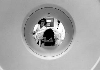
A research subject is positioned to enter NIDA's new state-of-the-art
PET scanner, used in these studies to detect and create images of brain
areas of increased glucose metabolism. Nurse Nelda Snidow draws blood samples
to monitor radioactivity levels.
The scanner, the cyclotron, and a radiochemistry laboratory are the key
components of the imaging center, which is funded in part by the White House
Office of National Drug Control Policy (ONDCP). General Barry R. McCaffrey,
director of the ONDCP, attended dedication ceremonies for the center along
with Dr. Harold Varmus, director of the National Institutes of Health (NIH);
Dr. Ruth L. Kirschstein, deputy director of NIH; NIDA Director Dr. Alan
I. Leshner; Dr. Barry J. Hoffer, director of NIDA's Division of Intramural
Research, and Center Director Dr. Edythe D. London.
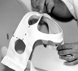 | Individually tailored masks are worn by research subjects
in the PET scanner. Dotted lines aid the technicians in precisely positioning
the subject's head inside the 360-degree core of the imager. |
Dr. London is a pharmacologist who has conducted innovative PET scan drug
abuse research, including mapping human brain areas involved in cocaine-induced
euphoria. Much of her earlier work used an older model PET scanner at Johns
Hopkins University in Baltimore. The Brain Imaging Center's scientific facility
and staff will be available to DIR scientists as well as to extramural researchers.
| The scanner can accurately depict images of physiological
features and activities with a resolution as small as 4 millimeters, which
is smaller than the size of a pea. |
 |
From NIDA NOTES, November/December, 1996
[NIDA Home Page][NIDA NOTES Index][1996 Archive Index Index]
|