Our Science – Austin Website
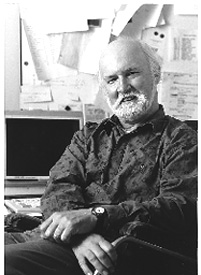
Stuart J. Austin, Ph.D.
 |
|
|||||||||||||||||||||||||
Biography
Dr. Austin received his Ph.D. from the University of Edinburgh in 1971 and was a postdoctoral fellow at Harvard Medical School and the University of Paris before joining the NCI-Frederick in 1976. He has been head of the Cell Cycle Regulation Section since 1984. In 1999, Dr. Austin joined the Center for Cancer Research, NCI.
Research
The Control of the Replication and Segregation of Bacterial Chromosomes. A fundamental property of cells is their ability to produce daughter cells that carry a faithful complement of genes. The mechanisms by which chromosome replication is controlled and the daughter chromosomes are equitably distributed to daughter cells are not understood in any detail. We seek to understand these processes at the molecular level. Because the problems are complex, we choose to tackle them by using the simplest possible model system. The bacterium Escherichia coli provides a powerful genetic system and, in addition to the bacterial chromosome itself, the organism provides a series of unique genetic elements that act as model chromosomes for some of our experiments. Extrachromosomal elements called unit-copy plasmids act as analogs of the chromosome and yet, unlike the host chromosome, are dispensable for cell survival. They are maintained at a 1:1 ratio with the host chromosome and are distributed with great precision to daughter cells. Our earlier studies focused on one of these elements, the plasmid prophage of bacteriophage P1. We continue these studies, but are now applying the tools we have developed and the information we have gained to try to understand how the host chromosome itself is organized and segregated.
The Role of Adenine Methylation in DNA Replication and Segregation. P1 DNA is controlled by a DNA marking system that distinguishes newly replicated DNA from unreplicated DNA and prevents a second round from occurring in a single cell cycle. Similar systems probably exist in all organisms, but the nature of the DNA marking and the replication block are generally not understood. Newly replicated P1 DNA is marked by hemimethylation of DNA sites within the origin of replication. The replication block is imposed by the recognition of the hemimethylated DNA by the SeqA protein, a protein whose selective DNA-binding activity was discovered in our laboratory. SeqA also regulates host chromosome replication in a similar fashion. However, we found that SeqA interacts with many other sequences than just the P1 and host chromosome origins of DNA replication. SeqA appears to interact with multiple hemimethylated GATC-binding sites to form a filament with the intervening DNA looped out. There are many potential sites distributed around the chromosome so that each replication fork should produce a transient wave of hemimethylation, and thus SeqA binding. Microscopy of cells with two replication forks showed pairs of SeqA-GFP foci located at the cell center. The number of foci increase sharply with increasing growth rate, corresponding approximately to the number of forks present in the cells. SeqA appears to organize newly replicated DNA into a structure that may aid DNA segregation away from replication forks that are tethered to the cell center.
The Roles of the Par and Muk Proteins in DNA Segregation. P1 has a DNA site, parS, that appears to be an analog of the centromere of eukaryotic chromosomes; parS promotes the accurate distribution (partition) of daughter plasmids to daughter cells. We have shown that the site is acted upon by two P1-encoded proteins that have been purified and characterized in our laboratory. The hypothesis that this par system is responsible for an activity akin to mitosis has recently received dramatic support. Homologs of the P1 ParA and ParB proteins have recently been shown to be responsible for the proper segregation of the chromosomes of two bacterial species. Moreover, studies of the location of these Par homologs within the cell and their likely DNA-binding sites provide important clues as to how partition works. Partition appears to involve attachment of chromosomes (or plasmids) to unique membrane-binding sites present in each half of the dividing cell. Condensation of the DNA then gathers each chromosome toward its point of attachment to allow cell division.
The E. coli MukB protein resembles eukaryotic dyneins, proteins that are involved in the energy-dependent movement of cell components. Mutants in the muk genes produce frequent anucleate cells. We discovered that mutations in topoisomerase I are general suppressors of muk mutants. These mutations eliminate anucleate cell production by increasing the negative supercoiling of the chromosomal DNA. This observation is consistent with a role for the Muk proteins in chromosome folding or DNA condensation. This result, along with our observations on the SeqA protein, support models in which chromosome replication is coreplicational. The newly replicated DNA emerges from the replication forks at the cell center and is directed toward each cell pole by membrane attachment and DNA condensation to form two separate chromosome masses.
Collaborators on this study have included Michael Davis and Anthony Maurelli, Uniformed Services University of the Health Sciences; Lyndsay Radnedge, Lawrence Livermore National Laboratory; Paul Tucker, European Molecular Biology Laboratory; and Andrew Wright, Tufts University.
This page was last updated on 6/11/2008.

