|
|
Letter
Echinococcus multilocularis
in Estonia
Epp Moks,* Urmas Saarma,*† and Harri Valdmann*
*University of Tartu, Tartu, Estonia; and †Estonian Biocentre, Tartu,
Estonia
Suggested
citation for this article
To the Editor: Alveolar echinococcosis (AE) caused by Echinococcus
multilocularis is one of the most important emerging zoonosis in Europe.
The fatality rate is >90% in untreated patients (1).
In Europe, the distribution range of the zoonotic tapeworm E. multilocularis
has expanded over the last few decades, and the parasite attracts increasing
awareness as a public health issue (2–5). In 2003, AE
was added to the list of zoonoses to be monitored in the member states
of the European Union, according to Directive 2003/99/EC.
This is the first report of E. multilocularis in Estonia, which
extends its northern distribution in Europe. Results of examinations of
17 red foxes shot in the eastern (Võnnu and Räpina) and western (Hiiumaa)
districts of Estonia from February to December 2003 were included in this
study. We examined the intestinal tracts by the sedimentation and counting
technique as described (1). Echinococcus adult
stages were found in 5 foxes (29.4%). Two foxes, infected with 3 and 5
adult worms, were from the Räpina district; 2 foxes, infected with 66
and 133 worms, were from the Võnnu district; and 1 fox, infected with
the highest number of worms (927), was from the Hiiumaa District. The
worms were retrieved, counted, washed, and stored in 90% ethanol until
DNA purification. The parasites were identified as E. multilocularis,
based on the most important morphometric parameters of adult stages (length
of worms, number of proglottids, terminal proglottids in percentage of
total worm length, position of genital pore, and form of uterus) (2).
To confirm the taxonomic status of the worms, polymerase chain reaction
(PCR) was conducted, followed by restriction fragment length polymorphism
(RFLP) analysis and direct sequencing of a portion of the NADH dehydrogenase
subunit I (ND1) gene of the mtDNA. A total of 6 specimens of E. multilocularis
were used for genetic analysis. Total genomic DNA was extracted with the
High Pure PCR Template Preparation Kit (Roche Molecular Biochemicals,
Mannheim, Germany) according to manufacturer's instructions. PCR-RFLP
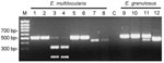
was performed as described by Gonzalez et al. (6). The
RFLP pattern of E. multilocularis isolates differed from that of
E. granulosus. Diagnostic cleavage at the locus Eg9 of E. multilocularis
with the enzyme CfoI is able to distinguish E. multilocularis
and its closest relative E. granulosus (Figure,
lanes 3 and 4 vs. lane 10). All 6 specimens of E. multilocularis
produced identical results. A 426-bp fragment of the mitochondrial ND1
gene was amplified with the primers NDfor2-AGTTTCGTAAGGGTCCTAATA and NDrev2-CCCACTAACTAACTCCCTTTC
using the BD Advantage 2 PCR Kit (Becton Dickinson Biosciences, Franklin
Lakes, NJ, USA) as described (7). DNA cycle sequencing
was performed by using the DYEnamic ET Terminator Cycle Sequencing Kit
(Amersham Pharmacia Biotech, Piscataway, NJ, USA). Sequences were resolved
on an ABI PRISM 377 automated DNA sequencer (Applied Biosystems, Foster
City, CA, USA).
All analyzed E. multilocularis specimens had identical sequences.
The ND1 sequence of E. multilocularis from Estonia was submitted
to GenBank under accession no. AY855918. The nucleotide sequences obtained
were compared with those in the GenBank sequence database. The sequence
of the Estonian isolate was identical with other E. multilocularis
sequences deposited under accession nos. AJ32907, AJ32908, AJ32909, and
AJ32910 from Poland (7) and AY389984 from China (Yang
JK et al., unpub. data), and differed considerably from the sequences
of the most closely related species, E. granulosus. For phylogenetic
analysis, the ND1 sequences of 7 E. multilocularis, 24 E. granulosus,
1 Taenia solium, 1 E. vogeli, and 1 E. oligarthrus
isolates were included and MrBayes 3.04b (8) was used
for the Bayesian estimation of phylogeny, applying the GTR+I+G substitution
model that best fitted the data (determined with Modeltest 3.06) (9).
Searches were conducted with 4 simultaneous Markov chains over 2 million
generations, sampled every 100 generations, and ended with a calculation
of a 50% majority rule consensus tree. On the phylogenetic tree, sequences
of Estonian isolate group together with those of other E. multilocularis
isolates from different countries and were clearly separated from those
of all other species (data not shown). The results of genetic analysis
confirmed morphologic identification of E. multilocularis.
This study reports a new location of E. multilocularis in Europe.
Estonia is the northernmost country on the mainland of the continent where
E. multilocularis has been described. Because no studies have been
published on the occurrence of E. multilocularis in Estonia in
either foxes or rodents, whether this report identifies a stable endemic
area or whether the parasite has expanded its range recently cannot be
determined. Although a limited number of foxes were examined, the occurrence
of E. multilocularis appears to be frequent and widespread in Estonia,
which poses a risk for putatively parasite-free adjacent countries in
Fennoscandia (2).
Acknowledgments
We thank Isam Sadula
Saeed for confirming the morphologic diagnosis of E. multilocularis.
Funding was provided
by Estonian Ministry of Education (target-financing grant 0181432) and
Environmental Investment Centre (target-financing grant 04-04-9/415).
References
- Kern P, Bardonnet K, Renner E, Auer H, Pawlowski Z,
Amman RW, et al. European
Echinococcosis Registry: human alveolar echinococcosis, Europe, 1982–2000.
Emerg Infect Dis. 2003;9:343–9.
- Eckert J, Gemmell MA, Meslin FX, Pawlowski ZS, editors. WHO/OIE Manual
on echinococcosis in humans and animals: a public health problem of
global concern. Paris: World Health Organization for Animal Health (Office
International des Epizooties) and World Health Organization; 2001.
- McManus DP, Zhang W, Li J, Bartley PB. Echinococcosis.
Lancet. 2003;362:1295–304.
- Sréter T, Széll Z, Egyed Z, Varga I. Echinococcus
multilocularis: an emerging pathogen in Hungary and Central Eastern
Europe? Emerg Infect Dis. 2003;9:384–6.
- Mazeika V, Paulauskas A, Balciauskas L. New data on the helminth fauna
of rodents of Lithuania. Acta Zool Lit. 2003;13:41–7
- Gonzalez LM, Daniel-Mwambete K, Montero E, Rosenzvit MC, McManus DP,
Carate T, et al. Further
molecular discrimination of Spanish strains of Echinococcus granulosus.
Exp Parasitol. 2002;102:46–56.
- Kedra AH, Swiderski Z, Tkach VV, Rocki B, Pawlowski J, Pawlowski Z.
Variability within NADH dehydrogenase sequences of Echinococcus multilocularis.
Acta Parasitol. 2000;45:353–5.
- Ronquist F, Huelsenbeck JP. MrBayes
3: Bayesian phylogenetic inference under mixed models. Bioinformatics.
2003;19:1572–4.
- Posada D, Crandall KA. MODELTEST:
testing the model of DNA substitution. Bioinformatics. 1998;14:817–8.
Suggested citation
for this article:
Moks E, Saarma U, Valdmann
H. Echinococcus multilocularis in Estonia [letter]. Emerg Infect
Dis [serial on the Internet]. 2005 Dec [date cited]. Available
from http://www.cdc.gov/ncidod/EID/vol11no12/05-0339.htm
|