|
||||||||||||||||||||||||||||||||||||||||||||||||||||||||||||||||||||||||||||||||||||||||||||||||||||||||||||||||||||||||||||||||||||||||||||||||||||||||||||||||||||||||||||||||||||||||||||||||||||||||||||||||
|
|
||||||||||||||||||||||||||||||||||||||||||||||||||||||||||||||||||||||||||||||||||||||||||||||||||||||||||||||||||||||||||||||||||||||||||||||||||||||||||||||||||||||||||||||||||||||||||||||||||||||||||||||||
Sexually Transmitted Diseases > Gonorrhea > Laboratory Information > Related
Species > Diplococcus mucosus
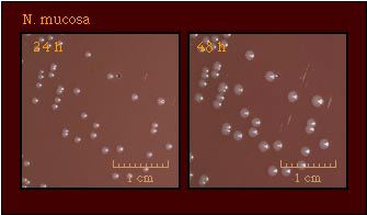
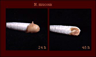
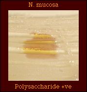
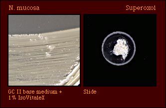
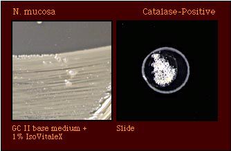
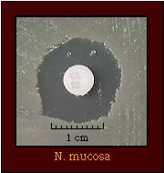
Diplococcus mucosus N. mucosa (Diplococcus mucosus) was described in 1906 but was not recognized again until 1959, when it was re-described by Veron et al. The failure to identify strains of N. mucosa occurred because nitrate reduction was not used as differential test until the 1960s. Strains of N. mucosa are distinguished from those of N. subflava biovar perflava and N. sicca by their ability to reduce nitrate. Table 1. Characteristics of N. mucosa
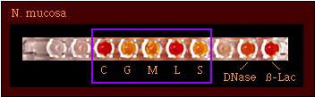
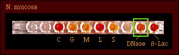
Species which may be misidentified as N. mucosa in acid detection testsThree Neisseria species, N. mucosa, N. sicca, and N. subflava biovar perflava, produce acid from glucose, maltose, fructose, and sucrose. N. mucosa may be identified by it's ability to reduce nitrate. Table 2. Characteristics of N. mucosa and other Neisseria spp. that produce acid from glucose, maltose, fructose, and sucrose
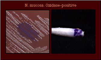
Abbreviations: G, glucose; M, maltose, S, sucrose; F, fructose; L, lactose; +, most strains positive; -, most strains negative; (+), some strains give weak positive reactions which are not representative of the species; R, strains grow well on selective medium for N. gonorrhoeae and/or show no inhibition around a colistin disk (10 micrograms); (R), most strains susceptible, some strains resistant. Although enzyme substrate tests are intended to be used only for the identification of Neisseria spp. isolated on selective media for N. gonorrhoeae, these tests do provide additional information that may aid in accurately identifying an isolate. However, N. mucosa produces prolyl aminopeptidase in enzyme substrate test and may be misidentified as N. gonorrhoeae if additional tests are not performed. Table 3. Supplemental tests which permit differentiation among Neisseria and related species that produce prolyl aminopeptidase in enzyme substrate tests
Abbreviations: G, glucose; M, maltose, S, sucrose; F, fructose; L, lactose; +, most strains positive; -, most strains negative; (+), strains may give weak positive reactions; R, strains grow well on selective medium for N. gonorrhoeae and/or show no inhibition around a colistin disk (10 micrograms); (R), most strains susceptible, some strains known to be resistant; S, all strains believed to be susceptible, no strains known to be resistant. ReferencesBovre K. Family VIII. Neisseriaceae Prevot, In N. R. Krieg (ed.). Manual of Systematic Bacteriology, vol. 1. The Williams & Wilkins co., Baltimore. 1984. p. 288-309. Knapp JS Historical perspectives and identification of Neisseria and related species. Clin Microbiol Rev 1988;1:415-431. Knapp JS, Rice RJ. Neisseria and Branhamella. In. Murray PR, Baron EJ, Pfaller MA, Tenover FC, Yolken RH. (ed.). Manual of Clinical Microbiology. 6th ed. American Society for Microbiology, Washington D. C, 1995. Vedros NA. Genus I. Neisseria Trevisan 1885, 105AL, In NR Krieg (ed.). Bergey's Manual of Systematic Bacteriology, vol. 1. The Williams & Wilkins Co., Baltimore. 1984. p. 290-296. von Lingelsheim W. Die bakteriologisch Arbeiten der Kgl. hygienischen Station zu Beuthen O.-Sch. wahrend der Genickstarreepidemie in Oberschlesien Im Winter 1904/05. Klin Jahrb 1906;15:373-489. Veron M, Thibault P, Second L. Neisseria mucosa (Diplococcus
mucosus Lingelsheim).
I. Description bacteriologique et etude du pouvoir pathogene. Ann Inst Pasteur
(Paris) 1959;100:497-500. Page last modified: October 17, 2008 Page last reviewed: October 24, 2008 Content Source: Division of STD Prevention, National Center for HIV/AIDS, Viral Hepatitis, STD, and TB Prevention |
||||||||||||||||||||||||||||||||||||||||||||||||||||||||||||||||||||||||||||||||||||||||||||||||||||||||||||||||||||||||||||||||||||||||||||||||||||||||||||||||||||||||||||||||||||||||||||||||||||||||||||||||
|
||||||||||||||||||||||||||||||||||||||||||||||||||||||||||||||||||||||||||||||||||||||||||||||||||||||||||||||||||||||||||||||||||||||||||||||||||||||||||||||||||||||||||||||||||||||||||||||||||||||||||||||||