|
Lab Manual Exercise # 1
Physical Properties & Structure of Cells
See The Cell & Mitosis Crossword Puzzle
If you have difficulty printing out this page, try the PDF version:
|
|
 |
Click PDF Icon To Read Page In Acrobat Reader. See Text In Arial Font Like In A Book.
View Page Off-Line: Right Click On PDF Icon To Save Target File To Your Computer.
|
Click Here To Download Latest Acrobat Reader. Follow The Instructions For Your Computer.
Section B. Onion Skin Cell
|
1. The cells of an onion skin are generally rectangular in shape and range in size from 0.25 to 0.4 millimeters in length (250-400 micrometers). A millimeter is abbreviated by mm and a micrometer by the Greek letter mu (12th letter of Greek alphabet) followed by an m:
|
|
millimeter
|
|
about 1/25th of an inch
|
|
micrometer
|
|
1/25,000th of an inch
|
|
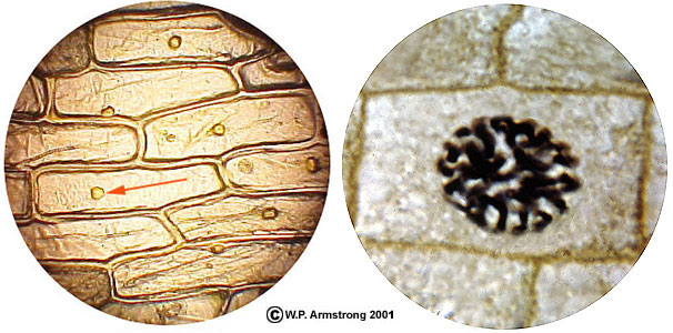
Left: Microscopic view of an onion skin showing several rectangular cells, each with a small, spherical nucleus (red arrow). The slide was stained with a drop of yellowish-brown gram's iodine. Right: Highly magnified view of a cell from the meristematic root tip of an onion showing enlarged nucleus containing 16 chromosomes. The cell is in prophase of mitosis, with distinct chromosomes (chromosome doublets) and a disintegrating nuclear membrane.
|
Diameter Of The Field Of View

|
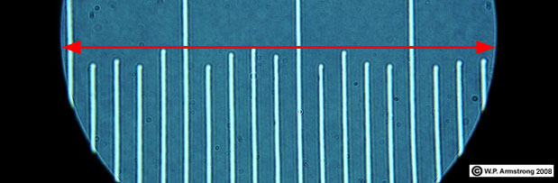
Compound microscope showing the 10x ocular (eyepiece) and four objectives (4x, 10x, 40x and 100x). [One objective is not in view.] To calculate the magnification, simply multiply the ocular lens (10x) by the objective lens. With this microscope you can obtain four different magnifications: 40x, 100x, 400x and 1000x.
|

|
The field of view when using the 10x objective (100x total magnification) is 2 mm. If 8 plant cells extend across the field of view (2 mm), then each cell is 2/8 or 0.25 mm long. Remember that the diameter of the field of view changes depending on the power of the objective according to the following table:
|
Objective
|
Diameter Of Field Of View
|
Magnification (10x Ocular)
|
4x
|
4.0 mm (4.45)
|
40x
|
10x
|
2.0 mm (1.78)
|
100x
|
40x
|
0.4 mm (0.45)
|
400x
|
100x
|
0.2 mm (0.178)
|
1000x
|
|
The original diameters of field of view (fov) were determined with a transparent mm ruler. This is like measuring the length of your finger nails with a yard stick. The values in parentheses are more precise. They were determined using a B & L stage micrometer.
If you know the diameter of the fov at one magnification, you can determine the diameter of fov at another magnification with the following formula:
diameter of fob#2 = diameter of fov#1 x magnification#1 divided by magnification#2
For example, if you know the diameter of fov at 100x magnification, the diameter of the fov
at 1000x magnification = 1.78mm x 100 divided by 1000 = 0.178 mm or 178 micrometers.
|
|
Stage micrometer at 1000x magnification with Olympus Compound Microscope. The diameter of field of view (fov) is 0.184 millimeters (184 micrometers). This corresponds to a 0.46 millimeter fov at 400 x magnification.
|
Relative Sizes Of Cells & Viruses 1
Diameter or Length
|
Millimeters
|
Micrometers
|
water molecule
|
0.000000385
|
0.000385
|
|
polio virus
|
0.00003
|
0.03
|
|
HIV retrovirus
|
0.0001
|
0.1
|
|
herpes virus
|
0.0002
|
0.2
|
|
Resolving Power of Average Light Microscope: 0.0003 mm
|
|
smallpox virus
|
0.0003
|
0.3
|
|
|
0.0005
|
0.5
|
|
|
0.0008
|
0.8
|
|
|
0.001
|
1.0
|
|
Staphylococcus bacterium
|
0.001
|
1.0
|
|
|
0.005
|
5.0
|
|
|
0.005
|
5.0
|
|
|
0.005
|
5.0
|
|
human red blood cell
|
0.0075
|
7.5
|
|
|
0.03
|
30
|
|
|
0.032
|
32
|
|
|
0.035
|
35
|
|
|
0.06
|
60
|
|
|
0.070
|
70
|
Resolving Power of Unaided Human Eye With 20-20 Vision
Able to focus on two hairs spaced 70 micrometers apart 8
|
|
|
0.085
|
85
|
|
|
0.20
|
200
|
|
|
0.30
|
300
|
|
Amoeba cell
|
0.30
|
300
|
|
|
0.30
|
300
|
|
|
0.40
|
400
|
|
|
0.60
|
600
|
|
|
1.50
|
1500
|
|
nerve cell (spinal cord)
|
600
|
600,000
|
|
|
100,000
|
100,000,000
|
|
|
1.3 x 10¹⁰
|
1.3 x 10¹³
|
|
1. The discovery of a virus called "mimivirus" in 1992 complicates the placement of viruses in the overall classification scheme for living organisms. Prior to this discovery, viruses were generally considered nonliving until they hijack a living cell. Officially, this virus got its name because it mimics bacteria (microbes) in size and complexity. It was placed in a new family of viruses, the Mimiviridae. Mimivirus was found inside an amoeba within a cooling tower in Bradford, UK. [The cooling tower was being investigated as the source of an influenza outbreak.] Mimivirus is the largest known virus, about 0.8 micrometers (800 nanometers) across. In fact it is larger than the bacterium causing gonorrhea.
|
Diameter of an Individual Mimivirus
|
|
metric unit
|
abbreviation**
|
decimal value
|
|
meter
|
m
|
0.000000800
|
|
millimeter
|
mm
|
0.000800
|
|
micrometer
|
µ
|
0.800
|
|
nanometer
|
nm
|
800
|
|
Angström
|
Å
|
8000
|
|
**html code for abbreviations may not show on all browsers.
|

|
With the exception of a some bacteriophages, viruses fall into two main morphological groups, those with cubical symmetry and those with helical symmetry. Until 1960, the only known examples of viruses with helical symmetry were those of plant viruses. The best studied example being the tobacco mosaic virus. The capsid denotes the protein shell that encloses the nucleic acid. It is composed of numerous repeating structural units. "Linear" viral capsids have RNA genomes that are encased in a helix of identical protein subunits. The length of the helical viral nucleocapsid is determined by the length of the nucleic acid.
|

|
Cubical viruses have the general molecular symmetry of an icosahedron. Although most viruses are not visible under an ordinary light microscope, mimivirus appears like a minute spheroid object under a compound microscope using an oil immersion objective (1000x magnification). An icosahedron has 20 identical equilateral triangles (facets), each subdivided into more equilateral triagular facets. There are electron micrographs of mimivirus available on the Internet. Simply do a search for mimivirus using one of the excellent search engines, such as google.com.
|
The mimivirus genome contains 1.2 million bases, more than many bacteria. The bases make up 1,260 genes, which makes it as complex as some bacteria. Most viruses use either DNA or RNA to carry their genetic information, but mimivirus has both of these nucleic acids. In addition, mimivirus can make about 150 of its own proteins, and can even repair its own DNA if it gets damaged. Normal viruses are not capable of protein synthesis or DNA repair on their own, they must rely on the organelles of their host cells for these activities. Whether mimivirus should be placed in an existing domain (superkingdom), or in its own domain, remains to be seen. For more information, see D. Raoult, et al. "The 1.2-Mb Genome Sequence of Mimivirus." Science Published On-line, DOI: 10.1126/Science.1101485 (2004); B. La Scola et al. "A Giant Virus in Amoebae." Science 299 (5615): 2033 (2003).
Some scientists have speculated that the evolution of eucaryotic cells involved the merging of two or more genomes, a phenomenon called symbiogenesis. Eukaryotic cells are characterized by the presence of membrane-bound organelles, including chloroplasts, mitochondria and nuclei. The origin of a complex cell has intrigued scientists for decades, and has been a strongly debated topic between evolutionists and creationists. According to the endosymbiont hypothesis, bacteria may be the progenitors of cellular organelles such as chloroplasts and mitochondria. A primitive nucleus (protonucleus) may have evolved from an intracellular virus; however, one weakness of this hypothesis is that viruses generally lack some of the key genes found in eukaryotes. The complex genome of mimivirus includes these genes, lending support to the evolution of a protonucleus from a virus.
Typical viruses are not placed in the three major domains of life. They are much smaller and much less complex than cells. They are macromolecular units composed of DNA or RNA surrounded by an outer protein shell. They have no membrane-bound organelles, no ribosomes (organelle of protein synthesis), no cytoplasm (living contents of a cell), and no source of energy production of their own. They do not exhibit autopoiesis--i.e. they do not have the self-maintenance metabolic reactions of living systems. Viruses lack cellular respiration, ATP-production, gas exchange, etc. However, they do reproduce, but at the expense of the host cell. Like obligate parasites, they are only capable of reproduction within living cells. In a sense, viruses hijack the host cell and force it to produce more viruses through DNA replication and protein synthesis. Outside of their host cells, viruses can survive as minute macromolecular particles. Viruses may attack animals and plants. Infectious human viruses can be dispersed though the air (airborne viruses) or body fluids (HIV virus). Epidemic viruses (such as HIV) that are passed from person to person via sexual conjugation are remarkably similar to computer viruses. Unfortunately in humans there is no resident antivirus program to alert you of a potential infection, or to quickly scan your body and delete the invader once it has entered your system. Humans must rely on their amazing antibody and cell-mediated immune responses, some of the most complex and remarkable achievements in the evolution of living systems.
2. This is the width of an oval (elliptical) spore. Spores of this size can easily escape from a folded (closed) paper envelope that is not airtight. Anthrax (Bacillus anthracis) is one of the microorganisms used in biological warfare because strains have been developed that are extremely infectious through the skin and through inhalation. In addition, it forms highly resistant spores that can survive for long periods. Approximately one teaspoon or two grams of anthrax may contain up to 20 billion spores. With an average infection rate of 10,000 spores per person, this is theoretically enough spores to infect 2 million people with inhalation anthrax.
|
Left: Highly magnified view (2000x) of human pus showing white blood cells (called neutrophils) with deeply-lobed purple nuclei. The minute paired dots (red arrow) are diplococcus gonorrhea bacteria (Neisseria gonorrhoeae). Each dot (coccus bacterium) is only about 0.5 micrometers in diameter. Some of the neutrophils have ingested the bacteria through phagocytosis. Right: A culture of rod-shaped anthrax bacteria (Bacillus anthracis). Some of the bacteria have divided by fission (red arrow). [Both images came from old (circa 1960) prepared microscope slides enhanced with Adobe PhotoShop by W.P. Armstrong.]
|
3. One human nucleus contains 46 chromosomes (single chromosomes during interphase). These 46 chromosomes comprise about 6 feet of DNA (2 meters). This amount of DNA was previously thought to contain about 100,000 functional genes, but that number has now been reduced to about 30,000 genes (one percent of the total DNA in the nucleus). In terms of information storage, the genetic information in a cell is roughly equivalent to 500 printed volumes of Encyclopedia Brittanica (12 characters per inch).
4. Mature sperm (spermatozoa) are composed of three distinct parts: a head, a middle piece (midpiece), and a tail (flagellum). The middle piece and tail contain microtubules in the characteristic 9 + 2 pattern of cilia and flagella (see animal cell illustration). In the middle piece, mitochondria are packed around the microtubules and provide the energy for movement. The journey of a single sperm in the female reproductive tract is analogous to a salmon swimming ten miles upstream to spawn. The head contains a nucleus covered by an outer cap called the acrosome which stores enzymes needed to penetrate the cellular and glycoprotein layers surrounding the egg.
Surgical sterilization of a man is called a vasectomy. Each of the two vas deferentia (sperm ducts) from the left and right testicles are cut and tied in two places, and the portion between the ties removed. Residual sperm may be present in the vas deferentia for up to a month following a vasectomy; therefore, periodic microscopic examinations are recommended until sperm are no longer visible in semen samplers.

|
Microscope image of human sperm in semen (1000x). The illustration insert shows the acrosome, head and middle piece of a human spermatozoan.
|

|
5. A baseball-sized puffball (Calvatia gigantea) in San Diego County, California. The inside is filled with a dark mass of spores intermingled with threadlike hyphae (capillitium). The close-up view of spores (right) was taken at 1,000x. Each spherical spore has a diameter of about 1/200 mm (5 µm), slightly smaller than a human red blood cell (7.5 µm). Depending on the species, puffballs range in size from a baseball to a basketball. When they
are mature, the puffball releases millions of spores into the air in a cloud of black dust. This can easily
be demonstrated if you accidentally kick one. According to David Arora (Mushrooms Demystified, 1986), a large puffball may contain 7 trillion spores. Lined up single
file, this number of spores would extend around the earth's equator. If each spore produced a
puffball the size of a basketball, the resulting puffballs would extend from the earth to the sun and
back!
|

|
6. Lichens are a symbiotic relationship between algae and fungi. The main body of a lichen is composed of fungal tissue (usually class Ascomycota) with photosynthetic algal cells (usually division Chlorophyta) embedded within this fungal mass (mycelium). The spores are produced by the fungal component or mycobiont, usually in a cup-shaped body called an ascocarp. Lichen spores can be 1-2 micrometers in Acarospora chlorophana, or up to 40 micrometers long in Diploschistes scruposus (left). The size, color and shape of spores are useful traits in separating different species of lichens. Spores up to 40 micrometers or longer are considered large.
|
|
7. The width (diameter) of a human hair ranges from very fine (0.017 mm) to very coarse (0.181 mm). The right image shows a fine human hair at 1,000 x magnification. The hair width is 0.070 mm or 70 micrometers. With the unaided eye the single hair shaft is visible when placed on a sheet of white paper.
|

|
8. A simple test for the resolving power of the unaided human eye:

|
A. Two fine human hairs are placed 70 micrometers apart. Each hair is approximately 70 micrometers in width (diameter). With 20-20 vision you should be able to differentiate between the two hairs at the proper focal distance. B. When placed closer to each other (about 15 micrometers apart), the two hairs appear to blend together and cannot be differentiated with the naked eye. A hand lens or magnifying glass is necessary to differentiate between the two hairs. Although the hairs can be seen on a white background, they cannot be resolved if they are placed close together. The minimum resolution for a computer monitor is 72 dots per square inch (dpi), otherwise the image becomes pixelated. Printed photos are also composed of dot patterns. Above 150 dpi, the dots generally blend together and cannot be resolved. Under a hand lens or microscope the dots can easily be observed.
|
9. Certain epiphytic orchids of the tropical rain forest produce the world's smallest seeds weighing only one 35 millionths of an ounce (1/35,000,000) or 0.81 micrograms. Some seeds are only about 1/300th of an inch long (85 micrometers). One seed capsule from a single flower may contain up to four million seeds. They are dispersed into the air like minute dust particles or single-celled spores, eventually coming to rest in the upper canopy of rain forest trees.
10. This minute one-seeded fruit is approximately 1/100 of an inch long and weighs about 70 micrograms or 1/400,000 of an ounce. Compare this tiny fruit with the most massive pumpkin that weighs over 1000 pounds (over 450,000 grams).
11. The plant body of the Australian species Wolffia angusta is only 0.6 mm long (1/42 of an inch). It weighs about 150 micrograms or 1/190,000 of an ounce. This is the world's smallest flowering plant, rivaled in minuteness by the Asian species W. globosa.
12. The Australian tree (Eucalyptus regnans) is the world's tallest flowering plant. Some of the largest trees are over 300 feet tall, rivaling in size the California coast redwood (Sequoia sempervirens).
13. The planet earth has a diameter of about 8,000 miles (13,000 kilometers) or 13 billion (13,000,000,000) millimeters. It has a volume of about one nonillion (1 X 1030) cubic millimeters. This astonishing volume is roughly equivalent to the volume of one nonillion wolffia plants packed together after 4 months of asexual budding!

|
If a water molecule is represented by 100, then a wolffia plant is about 1020 power larger than the water molecule. The earth is about 1020 power larger than a wolffia plant, or 1040 power larger than the water molecule.
|
|
Bird Eggs: World's Largest Cells By Volume
|
The typical bird egg contains an enlarged yolk surrounded by a protein-rich accessory fluid called the egg white or albumen. The yolk is essentially the enlarged ovum or egg cell. As this cell undergoes mitosis, the daughter cells become smaller and smaller until they are invisible to the naked eye. Egg-laying (oviparous) birds have large yolks rich in proteins and lipids to sustain the embryo. The developing chick obtains air through minute pores in the calcium carbonate egg shell. Pores in the shell are plugged by collagen, but are still permeable to air. Chicken egg shells can be brown or white, depending on the breed (variety). For example, Rhode Island Red, New Hampshire and Plymouth Rock varieties lay brown eggs. Most eggs purchased in your local market are unfertilized (haploid). Fertile (diploid) eggs are laid by hens regularly exposed to a rooster. The yolk contains a reddish-yellow carotenoid pigment (astaxanthin) which produces its bright color. In fact, chickens throughout the world are fed astaxanthin in their diet to intensify the yolk color.
A gigantic bird which lived in southern Madagascar until 900 AD produced the largest egg of any animal. Known as the elephant bird (Aepyornis maximus), it is an extinct member of the Rattae, a clade of flightless birds that includes the ostrich, emu, cassowary, kiwi and rhea, as well as the extinct moa of New Zealand. One enormous egg weighed about 27 pounds (13.6 kg) with a volume of 2.4 gallons (9 liters). This is roughly equivalent in size to 180 chicken eggs or 10,000 hummingbird eggs. The egg was much larger than any known dinosaur egg. It has been estimated that the elephant bird was ten feet (3 m) tall and weighed 1,000 pounds (450 kg). An egg preserved at the British Museum of Natural History measures 33.7 inches (85.6 cm) around the long axis, with a circumference of 28.5 inches (72.4 cm). The yolk of its egg was probably the largest single cell that ever existed. Unfortunately, the extinction of this magnificent bird was undoubtedly accelerated by early humans who invaded Madagascar.
The undisputed largest extant bird egg on earth today is laid by an ostrich. An average egg weighs about three pounds (1.4 kg), and is roughly equivalent to about two dozen chicken eggs. It would take approximately 40 minutes to hard-boil an ostrich egg. The yolk of an ostrich egg is the largest cell by volume; however, nerve cells from the spinal cord of a large hooved mammal may be nearly two feet (0.6 m) in length.
An egg case from a whale shark (Rhincodon typus) measuring 12 inches (30 cm) by 5.5 inches (14 cm) by 3.5 inches (9 cm) was discovered in the Gulf of Mexico in 1953 at a depth of 186 feet (56.6 m). The egg contained a perfect embryo of a whale shark 13.7 inches (35 cm) in length. This would certainly be the record for the largest extant embryo-containing egg; however, it would not be the largest single cell because it already divided into a multicellular embryo.
Viviparous animals with live birth and embryo development within the mother's uterus have minute eggs with very small yolks. There is no need for a large, nutrient-rich yolk because the embryo receives nutrients from the mother. Most mammals are viviparous, with the exception of the egg-laying duck-billed platypus. An unfertilized human egg (ovum) is roughly the size of a printed period at the end of this sentence. After fertilization it contains 46 chromosomes and approximately 30,000 functional genes, all the genetic information for a complete human organism. In terms of printed information using a 26 letter Roman alphabet and 12 characters per inch, this stored information represents about 500 volumes of Encyclopedia Brittanica.
|

|
The ostrich egg is the world's largest cell by volume. The actual cell portion is the inflated yolk (ovum) within the albumen (see chicken egg in following photo).
|

|
A chicken egg showing the yolk and white accessory fluid (albumen). The yolk is essentially the enlarged ovum or egg cell. The yellow color is enhanced by carotenoid pigments (astaxanthin) in the chicken feed. As this cell undergoes mitosis, the daughter cells become smaller and smaller until they are invisible to the naked eye. Egg-laying (oviparous) birds have large yolks rich in proteins and lipids to sustain the embryo. The nourishing albumen is also rich in proteins. During its embryonic development, the chick obtains air through minute pores in the calcium carbonate egg shell. Pores in the shell are plugged by collagen, but are still permeable to air.
|
Section B. Grains Of Salt & Metric System

|
Microscopic view of three cuboidal grains of ordinary table salt (sodium chloride or NaCl). All three grains are just over one millimeter in length (red bar). Grains of table salt vary slightly in size, but three average grains stacked together adds up to approximately one mm. If three grains equal one millimeter in length, then a single grain is approximately 0.3 mm or 0.03 cm on a side. Moving the decimal one place to the left converts millimeters (mm) to centimeters (cm).
|
|
How Much Does The Grain Weigh?
The volume of a single grain in cubic centimeters is (0.03)3 = 0.000027 cm3.
The density of NaCl is 2.165 grams per cubic centimeter. Multiply the density of NaCl by the volume of a single grain to obtain the weight of the grain:
2.165 g/cm3 x 0.000027 cm3 = 0.000058 g. This value can be rounded off to about 0.00006 g, the weight of one grain.
|
One grain weighs 0.00006 gram = 0.06 milligram = 60 micrograms
|
|
|
Although not precise (due to the variability in grains), the size and weight of a grain of table salt makes a nice comparison when discussing minute organisms or dosages. For example, the one-seeded fruit of Wolffia angusta and W. globosa (worlds smallest flowering plants) is roughly the size of a grain of table salt:
|

|
The minute one-seeded fruits of Wolffia angusta compared with grains of ordinary table salt (NaCl). The cubical salt grains are about 0.3 mm on a side. The fruits of W. globosa are similar in size.
|
|
One of the deadliest seeds on Earth is the castor bean (Ricinus communis). The seeds contain ricin, a very toxic protein compound known as a lectin. According to the Merck Index: An Encyclopedia of Chemicals, Drugs, and Biologicals (1997), a dose of ricin weighing only 70 micrograms or two millionths of an ounce (roughly
equivalent to the weight of a single grain of table salt from a salt shaker)
is sufficient to kill a 150 pound (68 kg) person. The colorful seeds of prayer bead (Abrus precatorius) contain another poisonous lectin called abrin. A dose of abrin equivalent to the weight of about 20 grains of table salt could be lethal to an adult human weighing 150 pounds. According to the Merck Index, one thoroughly masticated seed of prayer bead (A. precatorius) can cause fatal poisoning.
In 1978, a Bulgarian dissident, Georgi Markov, was assassinated in London after being pricked by a ricin-tipped umbrella. Ricin causes a slow and painful death through blood poisoning and a breakdown of the circulatory system. There is no known antidote for ricin poisoning. Even before the tragic terrorist plane crashes into the Trade Center Twin Towers in New York, some airports hand-inspected umbrellas packed in carry-on luggage. Following the Gulf War, UN investigator teams (UNSCOM) discovered that Iraq was purifying ricin for possible use in biological warfare, along with anthrax (Bacillus anthracis), botulism toxin (Clostridium botulinum), gas gangrene (C. perfringens), and aflatoxin (Aspergillus parasiticus). From: Facts on File News Services (23 January and 13 February 1998).
According to The Washington Post Online (16 November 2001), Osama bin Ladens's al-Qaida network also had working plans for making ricin. Instructions for preparing the poison were found in the cellar of an abandoned house once used as a terrorist training center. According to the article, the ricin may be ingested, injected and inhaled. The article also states that the laxative effect of castor oil is due to ricin, but this is doubtful. Castor oil is derived from the seeds of the castor bean. It contains 87 percent ricinoleic acid, a fatty acid with many industrial uses, along with small amounts of several other fatty acids including oleic (7%), linoleic (3%), palmitic (2%) and stearic (1%). The deadly protein ricin is not a component of purified castor oil.
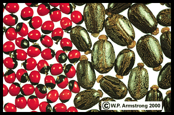
Seeds of prayer bead (left) and castor bean (right).
|
Section C. Elodea Leaf Cell
|
1. Depth of Field: When using a compound microscope this term refers to the thickness of the subject that is in focus. The Elodea leaf is composed of two layers of cells. Only one layer of cells is in focus when using the high power (40x) objective. Therefore, the depth of field is limited to only one layer of cells. You must focus up and down using the fine adjustment knob on the microscope to see both layers of cells.
2. Plasmolysis: Shrinkage of the protoplast or cell contents due to water loss when a drop of salt water (NaCl) is added to the Elodea slide. Because of all the salt ions (Na+ and Cl- ions) outside the cell membrane of each Elodea cell, water molecules move out of the cell membrane causing the cell membrane and it contents to shrink into a blob in the center of the cell wall. The porous, cellulose wall does not shrink because the salt ions easily pass through the wall but cannot pass through the membrane. Therefore, the cell membrane (not the cell wall) loses water molecules and shrinks.
|

|
Waterweed (Elodea densa) a submersed South American aquarium plant that is naturalized in ponds, streams and lakes throughout North America. It belongs to the frog's-bit family (Hydrocharitaceae) along with the troublesome Old World aquatic weed called hydrilla (Hydrilla verticillata) that literally clogs the waterways of canals and reservoirs. The highly magnified view of a leaf (right) shows a living photosynthetic cell. Because most of the cell is occupied by a water-filled, large central vacuole, the chloroplasts are displaced around the periphery of the cell, just inside the cell wall and membrane. The chloroplast shown by top red arrow is actually on the outside of this vacuole. Due to the limited depth of field at this magnification (400x), only portions of the viewing plane are in focus at any one time. To see additional chloroplasts you must focus up and down with the fine adjustment knob of the compound microscope. The transparent nucleus is not visible in this image. Please refer to the following illustration:
|

|
Elodea leaf cell illustration from a microscope slide. A drop of 10 percent NaCl (sodium chloride) solution was added to the slide. Left: The cell membrane has pulled away from the cell wall marking the onset of plasmolysis called "incipient plasmolysis." Right: The entire cell contents (protoplast) within the membrane has shrunk into a blob in the center of the cell. This phenomenon is called plasmolysis. Salt ions (Na+ and Cl-) have passed through pores in the cellulose cell wall. The ions do not pass through the protein-lipid cell membrane because it is impermeable to them. Due to the concentration differential inside and outside of the membrane, water molecules (shown by blue arrows) move out of the cell membrane, thus causing the cell contents to shrink into a blob. This concentration differential does not occur outside the porous, rigid cell wall, therefore its rectangular shape remains intact.
|

|
Microscopic view of the plasmolyzed cells of an Elodea leaf. A. Cell wall composed of cellulose. B. The contents (protoplast) of a cell that has shrunk into a ball after losing water. C. The faint cell wall of another layer of cells. Since only one layer of cells is in focus due to the depth of field at this magnification, the second layer of cells is blurry and faint.
|
Salt Intake & Decrease In Urine Output At A Movie Theatre
How To Sit Through a Long Movie, Such As Lord Of The Rings:
Two Towers, Without Leaving Your Seat To Drain Your Bladder.
- Visit the restroom during the latter part of the half-hour commercial/preview period, just before the movie starts.
- Do not drink diuretics, such as coffee or tea, three hours prior to the movie, and definitely not during the movie.
- Do not maintain your urine at a low specific gravity by drinking large quantities of water before or during the movie. In fact, it is wise to reduce your intake of all beverages during the movie.
- Eat salty popcorn during the movie. If it makes you thirsty, suck on some candy mints. As the salt content of extracellular fluids in the blood and kidneys goes up, so does the osmotic concentration and specific gravity of these fluids. Special sensors in the brain sense this change in salt concentration and stimulate the pituitary to secrete antidiuretic hormone (ADH). ADH is carried to the kidneys in the blood where it stimulates cells in the posterior convoluted tubules and the collecting ducts to reabsorb more water. In addition to returning more water to the blood, ADH also reduces sweating and causes constriction of arterioles. ADH is also referred to as vasopressin because it increases the blood pressure. These homeostatic actions of ADH result in decreased water loss and more concentrated urine. Consequently, your bladder does not fill as rapidly.
- Wear waterproof undergarments.
|
Important Definitions For The Cell Exam
|
1. Depth of Field: When using a compound microscope this term refers to the thickness of the subject that is in focus. The Elodea leaf is composed of two layers of cells. Only one layer of cells is in focus when using the high power (40x) objective. Therefore, the depth of field is limited to only one layer of cells. You must focus up and down using the fine adjustment knob on the microscope to see both layers of cells.
2. Plasmolysis: Shrinkage of the protoplast or cell contents due to water loss when a drop of salt water (NaCl) is added to the Elodea slide. Because of all the salt ions (Na+ and Cl- ions) outside the cell membrane of each Elodea cell, water molecules move out of the cell membrane causing the cell membrane and it contents to shrink into a blob in the center of the cell wall. The porous, cellulose wall does not shrink because the salt ions easily pass through the wall but cannot pass through the membrane. Therefore, the cell membrane (not the cell wall) loses water molecules and shrinks.
3. Hypertonic and Hypotonic: When comparing two solutions, such as the inside of a red blood cell (RBC) with the solution they are placed in, the solution with the greater salt concentration is hypertonic, while the solution with the lower salt concentration is hypotonic. Compared with the RBCs, a 10% NaCl solution is hypertonic while a 0.1 % NaCl is hypotonic. Since the latter solution is almost pure water, the blood cells are actually hypertonic by comparison.
4. Osmosis:
Movement of water molecules from a region of high concentration to a region of lower concentration through a differentially permeable cell membrane. In biology, this definition should include water molecules and cell membrane.
5. Diffusion: Movement of molecules or ions (charged atoms) from a region of high concentration to a region of lower concentration. This definition does not include a cell membrane and refers to molecules of any substance, not specifically water. Examples of diffusion include perfume molecules in the air, ether molecules in a classroom, sugar molecules in a cup of coffee, methylene blue molecules in a bowl of clear gelatin, etc.
6. Active Transport: Movement of molecules and ions through a cell membrane against a diffusion gradient (i.e. from a low to a higher concentration). This movement involves carrier proteins (channels) in the cell membrane and requires energy in the form of ATP (adenosine triphosphate). Active transport occurs during a nerve impulse or action potential (wave of depolarization) when a nerve cell membrane suddenly becomes permeable to sodium ions. The inflow of sodium ions moves quickly along the nerve cell axon, activating an adjacent nerve cell or muscle. As sodium ions rapidly move across the membrane to the inside of the axon, the polarity of the membrane changes. This reversal in polarity causes the sodium channels to close and the potassium channels to open. Now potassium ions move from inside the axon to the outside of the axon in a wave of repolarization.
During a lethal injection, a fatal dosage of potassium chloride is administered intravenously. The large influx of potassium ions interrupts the wave of depolarization (action potential) to the heart muscle resulting in cardiac arrest. A paralyzing agent, such as pancuronium bromide or tubocurarine chloride, plus a lethal dosage of a general anesthetic, such as sodium thiopental (sodium pentothal) are usually given before the potassium chloride is administered. Tubocurarine is the active ingredient of curare, an extract from the bark and stems of the South American vine (Chondodendron tomentosum). Amazonian Indians use the gummy extract to coat the poison darts of their blowguns. The alkaloid D-tubocurarine blocks acetylcholine receptor sites at neuromuscular junctions, causing relaxation and paralysis of muscles, including respiratory organs and the heart.
6. Halophyte: A plant adapted to soil or water containing a high salt concentration. The root cells contain a salt concentration higher than the water they are growing in. Since they are hypertonic compared with the water or soil, the root cells can take in water molecules and do not become plasmolyzed. Good examples of halophytes are the seagrasses (see link below), red and black mangroves (Rhizophora and Avicennia), salt grass (Distichlis) and salt bush (Atriplex). There are also species of bacteria and algae that are extremely halophilic (salt-loving), living in saturated brine and salt crust (see link below).
7. Imbibition: The movement of water molecules into a porous, colloidal substance causing it to swell. Enormous pressures can be produced by imbibition, sufficient to split boulders apart.

|
When a seed of Sterculia lychnophora is soaked in water for several days, it imbibes water and swells to more than eight times its original volume. The seed coat expands into an edible gelatinous mass (carbohydrate gum) that is used for a beverage in Cambodia. According to S. Facciola (Cornucopia II, 1998), the cooling beverage is called "sam-rong," and the flavor is enhanced with sugar and a flavoring such as jasmine or banana water.
|
8. Light Reactions of photosynthesis: The production of ATP and NADPH2 within the grana region (thylakoid membranes) of a chloroplast using light energy.
9. Dark Reactions of photosynthesis (also known as the Calvin Cycle): The conversion of carbon dioxide into glucose within the stroma region of a chloroplast. This complex series of reactions requires ATP and NADPH2 from the light reactions.
10. Fluorescence: The emission of a reddish glow when a beam of light is directed on a chlorophyll solution.
11. Bioluminescence: The emission of light by an organism or population of organisms. This phenomenon involves the oxidation of luciferin in the presence of ATP and the enzyme luciferase. Examples of bioluminesence include dinoflagellates causing "red tide," lightning "bugs" (beetles), glow worms (beetle larvae), comb jellies (phylum Ctenophora), the deep sea angler fish, and a remarkable fungus called the jack-o-lantern mushroom.
|
Section D. Potato Tubers & Banana Fruits
|
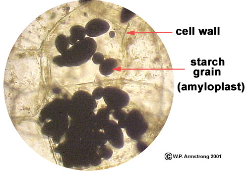
The thin-walled parenchyma cells of a potato tuber are filled with membrane-bound, starch-storage organelles called amyloplasts. They are also referred to as "starch grains" in most general biology textbooks. Since iodine stain (gram's iodine) makes starch turn purplish-black, the amyloplasts can easily be viewed with a compound microscope (400x). Insoluble starch (amylopectin) is deposited in concentric layers within the amyloplasts. Unlike the long, coiled molecules of soluble starch (amylose), the molecules of amylopectin are much shorter, with only 40-60 glucose subunits. Amylopectin molecules consist of highly branched chains that do not coil. Starch grains of different plant species have characteristic shapes, such as maize (corn), oats, bananas, potatoes and wheat. For example, banana starch grains are more elongate than potato starch grains. Starch is hydrolyzed (broken down) by amylase enzymes (including B-amylase and maltase). During hydrolysis a water molecule is inserted between each glucose subunit. Starch is typically stored in underground organs, including storage roots, rhizomes, tubers, corms and bulbs.
|
Magnified view (400x) of several parenchyma cells of a potato tuber showing the thin, transparent cell walls and clusters of amyloplasts (starch grains). The starch grains were stained black with gram's iodine.
|
|
Section E. Oak Wood & Specific Gravity
|
1. As a tree grows in girth, the marked difference in the size and density of spring and summer cells produces concentric annual rings. Because the summer wood cells in pine trunks are smaller and more dense, they appear as dark bands in a cross section of a log. Each concentric band of spring and summer cells is called an annual ring. By counting the rings (dark bands of summer cells in pine wood), the age of the tree can be determined. Other data, such as fire and climatic data, can be determined by the appearance and spacing of the rings. Some of the oldest bristlecone pines (Pinus longaeva) in the White Mountains of eastern California have more than 4,000 rings. Annual rings also produce the characteristic grain of the wood, depending on how the boards are cut at the saw mill.
2. To calculate the specific gravity of a block of wood, divide the numerical value for its weight by the numerical value for its volume. Specific gravity is expressed as a number, without any units of measurements. For more information about this subject, refer to the Wayne's Word article about hardwoods.
Specific Gravity
Probably the best way to appreciate the relative hardness of different woods is the concept of "specific gravity," a numerical scale based on 1.0 for pure water. Without getting too mathematical, the specific gravity of a substance can easily be calculated by dividing its density (in grams per cubic centimeter) by the density of pure water (one gram per cubic centimeter). The brilliant Greek mathematician and inventor Archimedes discovered over 2,100 years ago that a body in water is buoyed up by a force equal to weight of the water displaced. Archimedes reportedly came upon this discovery in his bathtub, and ran out into the street without his clothing shouting "Eureka, I have found it." Since one gram of pure water occupies a volume of one cubic centimeter, anything having a specific gravity greater than 1.0 will sink in pure water. The principles of buoyancy and specific gravity are utilized in many ways, from scuba diving and chemistry to the hardness of dry, seasoned wood. Some of the heaviest hardwood trees and shrubs of the United States have specific gravities between 0.80 and 0.95; including shagbark hickory (Carya ovata), persimmon (Diospyros virginiana) and ironwood (Ostrya virginiana) of the eastern states, and canyon live oak (Quercus chrysolepis), Engelmann oak (Q. engelmannii), hollyleaf cherry (Prunus ilicifolia) and Santa Cruz Island ironwood (Lyonothamnus floribundus ssp. asplenifolius) of southern California. Although some of these trees are called ironwoods, their dense, dry wood will still float in water. Since the pure cell wall material (lignin and cellulose)) of wood has a density of about 1.5 grams per cubic centimeter, even the world's heaviest hardwoods generally have specific gravities less than 1.5 due to tiny pores (lumens) within the cell walls. True ironwoods include trees and shrubs with dry, seasoned woods that actually sink in water, with specific gravities greater than 1.0. They include lignum vitae (Guaicum officinale, 1.37); quebracho (Schinopsis balansae, 1.28); pau d'arco (Tabebuia serratifolia, 1.20); knob-thorn (Acacia pallens, 1.19); desert ironwood (Olneya tesota, 1.15); and ebony (Diospyros ebenum, 1.12). To appreciate the weight of these hardwoods, compare them with tropical American balsa (Ochroma pyramidale), one of the softest and lightest woods with a specific gravity of only 0.17.
|
|
Section F. Cheek Squamous Epithelial Cells

|
Magnified view (400x) of squamous epithelial cells from the buccal mucosa (cheek cells from inside the mouth). The cells are stained with a dye called methylene blue. The nucleus and cell membrane are clearly visible. Plant structures such as a cell wall, chloroplasts and large central vacuole are absent. Because they don't have a rigid (firm) cellulose cell wall, these cells are flimsy and irregular in shape, unlike the rectangular shape of the onion cells. Although they were photographed in fall of 2001, these cells actually came from a student in a previous biology lab three years before.
|
Section G. Generalizations
|
1. The cells of eukaryotic plants, protists, and animals are basically similar because they have many structures in common. They both have a cell membrane, nucleus and a cytoplasm composed of many of the same organelles. The term cytoplasm refers to the region of a cell outside of the nucleus. Some of the organelles they have in common are ribosomes (site of protein synthesis), mitochondria (site of cellular respiration and ATP production), Golgi apparatus (vesicles involved in the secretion of macromolecules from cells), and lysosomes (vesicles involved in the intracellular digestion of macromolecules). In addition, they both have complex networks of intracellular canals called the endoplasmic reticulum through which macromolecules move.
2. Three obvious characteristics of plant cells that are not found in typical animal cells are: a cellulose cell wall, a large central vacuole, and chloroplasts (site of photosynthesis).
3. The cells within an organism are basically similar. They differ in their size, shape and function. Cells are organized into tissues (nerve, muscle, adipose, epithelial, etc.) and tissues are organized into organs (heart, liver, brain, kidneys, etc.). Although they are not as anatomically complex, plants also have organs (leaf, root, flower, etc.). Plants probably equal or exceed the complexity of animals when it comes to their incredible number of diverse biochemical reactions and products.
|
Section H. Prokaryotic Cells
|
1. Nitrogen Fixation: A remarkable
prokaryotic skill in which inert atmospheric nitrogen gas
(N2) is combined with hydrogen to form ammonia
(NH3). This process occurs in the root nodules of legumes, and is the main reason why farmers often rotate their crops with leguminous species (such as alfalfa). This process also occurs in a number of species of microscopic cyanobacteria, some of which live symbiotically in the leaves and roots of plants. The actual sites of nitrogen fixation in the cyanobacteria are special cells called heterocysts.
Unlike other cells in the filaments of cyanobacteria, the heterocyst is nonphotosynthetic. As the heterocyst matures, the photosynthetic membranes (thylakoid membranes) become contorted or reticulate compared to regular photosynthetic cells of cyanobacteria, and
they become non-photosynthetic (and do not produce oxygen). This
fact is especially noteworthy because nitrogen fixation requires
the essential enzyme nitrogenase, and the activity of
nitrogenase is greatly inhibited by the presence of oxygen.

|
Filamentous cyanobacteria (Anabaena azollae) live inside cavities within the leaves of the ubiquitous water fern (Azolla filiculoides). The larger, oval cells are heterocysts (red arrow), the site of nitrogen-fixation where atmospheric nitrogen (N2) is converted into ammonia (NH3). Polar nodules are visible in some of the heterocysts. The water fern benefits from its bacterial partner by an "in house" supply of usable nitrogen. The cellular structure of these bacteria has changed very little in the past one billion years.
|
2. Nitrification: A prokaryotic skill in which the ammonia from nitrogen fixation is converted into nitrites (NO2-) and nitrates (NO3-).
3. Ammonification: A prokaryotic skill in which ammonia is formed from the decay of protein.
Although our atmosphere is almost 80% nitrogen gas (N2), the element nitrogen is unavailable to plants in the inert gaseous state. Nitrogen fixation, nitrification, and ammonification make nitrogen available to autotrophic plants and ultimately to all members of the ecosystem. Symbiotic relationships such as the water fern (Azolla) are especially interesting because this little aquatic fern obtains a rich supply of nitrogen in the form of ammonia from the cyanobacteria (Anabaena)
living within cavities in its leaves.
4. Denitrification: A prokaryotic skill in which nitrites and nitrates are converted back into unusable, inert, nitrogen gas by bacteria in the soil and water. Luckily for plants and animals on the earth, natural nitrogen fixation exceeds denitrification. However, the serious problem today is that humans have greatly accelerated nitrogen fixation by the tremendous production of chemical fertilizers. These fertilizers eventually get into lakes and ponds resulting in excessive growth (blooms) of filamentous algae which form scummy masses on the water surface. Although algae produce oxygen through photosynthesis, the decay (oxidation) of these massive algal blooms actually depletes the oxygen supply in the water, resulting in massive fish kills and putrefaction of lakes and ponds. The biological term for this process is called eutrophication. Although eutrophication is a natural process it has been accelerated by humans to an alarming rate. It is even beginning to be noticeable in clear mountain lakes, such as Lake Tahoe, where the water is not quite as clear.
5. The three major types of archaebacteria are: (1) Methanogens (methane-producers) which are responsible for swamp gas; (2) Extreme Thermophiles that live in hot springs and black smokers (heat vents) at the bottom of the ocean; and (3) Extreme Halophiles (halobacteria) that live in saturated brine and salt crust.
6. The archaebacteria have some characteristics that are very different from the true bacteria (eubacteria) and cyanobacteria in the kingdom Monera. Lipids of archaebacterial cell membranes differ considerably from those of both prokaryotic and eukaryotic cells, as do the composition of their cell walls and the sequence of their ribosomal RNA subunits. In addition, recent studies have shown that archaebacterial RNA polymerases resemble the eukaryotic enzymes, not the eubacterial RNA polymerase. Some authorities hypothesize that eukaryotic organisms may have evolved from ancient archaebacteria (archae = ancient) rather than from the common and cosmopolitan eubacteria. The archaebacteria could have flourished more than 3 billion years ago under conditions previously thought to be uninhabitable to all known life forms. Although many conservative references place the archaebacteria in a separate division within the kingdom Monera, some authorities now recognize them as a new 6th kingdom--The kingdom Archaebacteria. [Some references state that the genes of archaebacteria are edited before they are translated into protein, a complex pathway known to occur in eukaryotic cells; however, more scholarly references do not mention this DNA similarity with eukaryotic cells, so it will not be postulated here.]
The vacuoles of some members of the duckweed family (Lemnaceae) contain calcium oxalate crystals that are visible under 400x magnification. Crystals of calcium oxalate may be needle-like (raphide crystals) or many faceted like a glistening diamond (druse crystals). The raphide crystals in the plant body of Lemna species appear like minute microscopic needles. Needlelike raphide crystals are also found in members of the arum family (Araceae), including the common house plant called Dieffenbachia. The crystals may cause irritation and swelling of the tongue if you chew on the leaves of this plant. Comparative DNA studies have shown that arums are closely related to the duckweed family. For more information about this remarkable family of flowering plants, refer to the following link:
|
|


