 |
 |
|
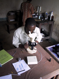
Migrant workers clinic at Mawker Tai camp in Thailand near the Myanmar
border. Microscopy plays a critical role in diagnosis of malaria,
one of the major health problems in this area. Image contributed
by the Shoklo Malaria Research Unit, Mae Sot, Thailand.
|
Microscopic
examination remains the "gold standard" for laboratory confirmation
of malaria.
Technique
(See DPDx
specimen preparation) A blood specimen collected from the patient
is spread as a thick or thin blood smear, stained with a Romanovsky
stain (most often Giemsa), and examined with a 100X oil immersion
objective. Visual criteria are used to detect malaria parasites and
to differentiate (when possible) the various species. (See DPDx
Plasmodium species comparison chart)
Advantages
Microscopy is an established, relatively
simple technique that is familiar to most laboratorians in endemic countries.
In such areas, microscopy is a standard technique used for diagnosing
other diseases (such as tuberculosis), often by the same laboratorians
using the same facilities and equipment.
Disadvantages
In many developing countries, microscopy
is not reliable because the microscopists are insufficiently trained
and supervised and are overworked, the microscopes and reagents are of
poor quality, and often the supply of electricity is unreliable. Conversely
in non-endemic countries, laboratory technicians are often unfamiliar
with malaria and may miss the parasites.
 |
|
Laboratory technician in Tanzania performing microscopic diagnosis.
Such work is often conducted under difficult conditions. The illumination
source is a portable, battery-powered device. |
Addressing
The Problems of Microscopy
Page last modified : September 14,
2006
Content source: Division of Parasitic Diseases
National Center for Zoonotic, Vector-Borne, and Enteric Diseases (ZVED)
|
|
|