Scientists Observe Infectious Prion Proteins Invade and Move Within Brain Cells
Scientists for the first time have watched agents of brain-wasting diseases, called transmissible spongiform encephalopathies (TSE), as they invade a nerve cell and then travel along wire-like circuits to points of contact with other cells. These findings will help scientists better understand TSE diseases and may lead to ways to prevent or minimize their effects. TSE, or prion, diseases include scrapie in sheep and goats; chronic wasting disease in deer and elk; mad cow disease in cattle; and Creutzfeldt-Jacob disease in humans.
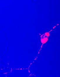 |
Prion trafficking in nerve cells. Prions branded with a fluorescent dye (pink) were added to nerve cells taken from a hamster brain. The prions initially were in the form of large clumps on the cell, but over time the clumps were broken into smaller units and transported along wire-like nerve cell projections. |
Under the direction of Byron Caughey, Ph.D., at the Rocky Mountain Laboratories (RML), and Marco Prado, Ph.D., at the University of Minas Gerais in Belo Horizonte, Brazil, the team performed the research in laboratory cultures using a rodent-adapted form of scrapie protein and cells taken from the central nervous system of mouse and hamster brains. The proteins were first “branded” with fluorescent dyes so they could be easily tracked. View prion trafficking in nerve cells.
The work also revealed that a similar trafficking process might occur with the key plaque-forming protein in Alzheimer’s disease, which is not a TSE but a different type of degenerative brain disease, according to Gerald Baron, Ph.D., one of the lead RML researchers. RML, located in Hamilton, MT, is part of the National Institute of Allergy and Infectious Diseases (NIAID) of the National Institutes of Health. The new report appears in the May 25 issue of The Journal of Neuroscience.
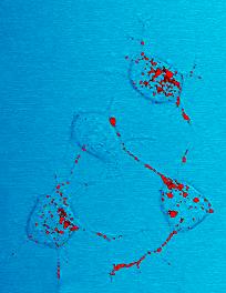 |
| Prions branded with a fluorescent dye (red) were added to nerve cells taken from mice. The prions initially were in the form of large clumps on the cell, but over time the clumps were broken into smaller units and transported along wire-like nerve cell projections. |
“These findings offer intriguing leads toward developing therapies to stop the spread of TSE and possibly other degenerative brain diseases,” says NIAID Director Dr. Anthony Fauci. “Potentially, it may be possible to block the pathways that prions use to invade cells, their exit to other cells or their replication within the cells.”
Those are precisely some of the next experiments the RML group is pursuing, along with trying to move the fluorescent tracking method from laboratory cell cultures to live mice and hamsters. Along with Drs. Caughey, Prado and Baron, other key researchers involved in the project included Kil Sun Lee, Ph.D., RML, and former RML employee Ana Cristina Magalhães, Ph.D., also from the Federal University of Minas Gerais.
Dr. Baron explains that throughout his seven years at RML, he and others have contemplated how to use fluorescent tracking to learn more about TSEs, but they struggled to develop an effective method to do so.
“When I started working on TSEs, I thought about them as being similar to intracellular bacterial pathogens—something that replicates within an animal or human host cell,” says Dr. Baron. “I wanted to know how such a pathogen binds to the host cell, and how it enters, replicates and spreads to other cells.”
Dr. Baron says researchers have tracked infectious prion protein moving through other parts of animal bodies up to the brain, but no one had ever tracked the protein movement within animal brain cells. One of the most difficult aspects of the experiment, he says, was finding a way to fluorescently tag the TSE prion proteins without altering them—while still allowing researchers to identify the prions as they penetrated the cells and spread within the long projections that nerve cells develop to send signals to other nerve cells.
“This was difficult from a technical aspect because the scrapie pathogen is largely a corrupted form of a host cell protein,” Dr. Baron said. “It can be hard to detect the corrupted prion protein in living infected cells and distinguish it from its normal counterpart.”
He explains that once researchers learned how to mark the prion proteins, they added them to a culture of nerve cells and then began watching and taking photo images with a confocal microscope. Confocal microscopy uses laser light to scan many thin sections of a fluorescent sample, resulting in a clean three-dimensional image. The painstaking job of analyzing and deciphering about 1,000 different images primarily belonged to Dr. Magalhães—who filled a file cabinet drawer with CDs containing microscopic images. The effort resulted in striking photos that, when put into a video format, show prion protein moving within cells, then along narrow cellular projections called neurites and ultimately into close proximity with adjacent cells.
Other areas the research group plans to explore include
- Exactly where within nerve cells does scrapie infection occur, and how does this happen?
- How and why do large masses of infectious prion protein attach to host cells and become broken into smaller units so that they can invade the cell interior?
- What types of chemical messages are sent between neurites from one cell to another that allow infectious prions to transfer between cells?
- What happens to the infectious prion protein once it is transferred to another cell?
- How do the many different possible pathways that lead into cells determine what happens to prion protein; some pathways could lead to digestion by the cell, others lead to transfer—and presumably infection—in adjacent cells.
“This has been pretty amazing—certainly a new approach for our field,” Dr. Baron says.
NIAID is a component of the National Institutes of Health, an agency of the U.S. Department of Health and Human Services. NIAID supports basic and applied research to prevent, diagnose and treat infectious diseases such as HIV/AIDS and other sexually transmitted infections, influenza, tuberculosis, malaria and illness from potential agents of bioterrorism. NIAID also supports research on transplantation and immune-related illnesses, including autoimmune disorders, asthma and allergies.
###
Reference: A Magalhães et al. Uptake and neuritic transport of scrapie prion protein coincident with infection of neuronal cells. The Journal of Neuroscience 25 (21):5207-16 (2005). DOI: 10.1523/JNEUROSCI.0653-05.2005.
NIAID is a component of the National Institutes of Health (NIH), an agency of the U.S. Department of Health and Human Services. NIAID supports basic and applied research to prevent, diagnose and treat infectious diseases such as HIV/AIDS and other sexually transmitted infections, influenza, tuberculosis, malaria and illness from potential agents of bioterrorism. NIAID also supports research on transplantation and immune-related illnesses, including autoimmune disorders, asthma and allergies.
back to top