|
|
Dispatch
Domestic Poultry and SARS
Coronavirus, Southern China
David E. Swayne,* David L. Suarez,* Erica Spackman,* Terrence M. Tumpey,* Joan R. Beck,*
Dean Erdman,† Pierre E. Rollin,† and Thomas G. Ksiazek†
David L. Suarez,* Erica Spackman,* Terrence M. Tumpey,* Joan R. Beck,*
Dean Erdman,† Pierre E. Rollin,† and Thomas G. Ksiazek†
*U.S. Department of Agriculture, Athens, Georgia, USA; and †Centers for
Disease Control and Prevention, Atlanta, Georgia, USA
Suggested citation
for this article:
Swayne DE, Suarez DL, Spackman E, Tumpey TM, Beck JR, Erdman D, et al.
Domestic poultry and SARS coronarvirus, southern China. Emerg Infect
Dis [serial online]. 2004 May [date cited]. Available from: http://www.cdc.gov/ncidod/EID/vol10no5/03-0827.htm
SARS coronavirus
injected intratracheally into chickens, turkeys, geese, ducks, and quail,
or into the allantoic sac of their embryonating eggs, failed to cause
disease or replicate. This finding suggests that domestic poultry were
unlikely to have been the reservoir, or associated with dissemination,
of SARS coronavirus in the animal markets of southern China.
An outbreak of severe acute respiratory syndrome (SARS) occurred in Guangdong
Province, People’s Republic of China, in November 2002 and spread to patients
in 30 countries in Africa, Asia, Australia, Europe, and North and South
America (1,2). As of July 11, 2003, SARS had been diagnosed
in 8,437 patients; 813 died (1). A novel coronavirus
was isolated in tissue culture or detected by reverse transcription–polymerase
chain reaction (RT-PCR) from multiple respiratory specimens in many patients
with SARS (2–4). The SARS-coronavirus (SARS-CoV) is proposed
to be the cause of this syndrome on the basis of its association with
human clinical cases (3,4) and reproduction of pulmonary
lesions in experimentally challenged cynomolgus macaque monkeys (Macaca
fascicularis) (5). Furthermore, some of the first
persons identified with SARS-CoV infections were vendors in animal markets
of southern China, which suggests a possible animal source (6).
SARS-CoV has been detected by real-time RT-PCR or isolated from two wild
mammalian species, Himalayan palm civet (Paguma larvata) and raccoon
dog (Nytereutes procyonoides), in a market in southern China (7),
but other studies in southern China involving six provinces and Beijing,
as well as sampling of 54 wild and 11 domestic animal species, did not
find SARS-CoV (8). The original source of this virus
remains unknown (3). The susceptibility of different
animal species within the animal meat markets is unknown.
Coronaviruses have been identified in numerous mammalian and avian hosts.
Most widely studied and of common occurrence are coronaviruses reported
in chickens (infectious bronchitis virus), turkeys (turkey enteric coronaviruses),
cats (feline infectious peritonitis virus and feline enteric coronavirus),
dogs (canine enteric coronaviruses), swine (porcine hemagglutinating encephalomyelitis
virus, porcine transmissible gastroenteritis virus, and porcine respiratory
coronavirus), cattle (bovine enteric and respiratory coronaviruses), mice
(Murine hepatitis virus), rats (sialodacyradenitis virus), rabbits (rabbit
coronavirus), and humans (respiratory and enteric coronaviruses) (9).
However, on the basis of sequence data, SARS-CoV is sufficiently different
from these known group 1, 2, and 3 animal and human coronaviruses to be
classified as a new group, group 4 coronaviruses (10).
Most likely SARS-CoV originated from an unknown animal reservoir, not
from a benign coronavirus in the human population (10,11).
Domesticated poultry species are major commodities traded in the animal
markets of southern China. Poultry have been shown to be reservoirs for
H5N1 and H9N2 avian influenza viruses that have crossed over and caused
infections in humans from 1997 to 2003, some with fatal outcomes (12–14).
Therefore, poultry should be examined as potential hosts for infection
and amplification of SARS-CoV to determine any potential role they may
have played during the emergence of human infections in southern China.
Groups of nine 3-week-old domestic geese (Anser anser domesticus),
3-week-old domestic Pekin ducks (Anas platyrhyncos), 4-week-old
chickens (Gallus gallus domesticus), 3-week-old turkeys (Meleagris
gallopavo), and 5-week-old Japanese quail (Coturnix coturnix japonicus)
were each injected intratracheally with 106.2 mean tissue culture
infective doses (TCID50) of Vero E6 propagated Urbani SARS-CoV
per bird in a volume of 0.1 mL. The inoculum was the third passage in
Vero E6 cells from the original throat swab specimen of the patient. The
chickens were specific pathogen–free from an inhouse flock. The other
four species were conventional birds obtained at 1 day (geese, turkeys,
and ducks) or 5 weeks of age (quail) from commercial hatcheries and raised
on site. Oropharyngeal and cloacal swabs were obtained on days 0, 1, 2,
3, 4, and 10 after injection from five birds per group for virus detection
by real-time RT-PCR and virus isolation on Vero E6 cells. RNA for RRT-PCR
was extracted with the Trizol LS reagent (Invitrogen, Carlsbad, CA) in
accordance with the manufacturer's instructions. Two hydrolysis probe
type real-time RT-PCR assays, both targeting the ORF 1b gene, were optimized
and run on a Smart Cycler (Cepheid, Sunnyvale, CA) with the superscript
platinum taq one-step RT-PCR kit (Invitrogen, Carlsbad, CA). Real-time
RT-PCR tests included negative (noninfected tissue culture media, infectious
bronchitis coronavirus, and turkey enteric coronaviruses) and positive
(Vero E6 propagated SARS-CoV) controls. Two injected birds of each species
were euthanized. After necropsy, their tissues were collected for histopathologic
examination (all tissue types) and virus detection (plasma, trachea, lung,
spleen, kidney, and heart) on days 2 and 4 after injection, and at termination
on day 10 after injection. For determination of infection, serum was collected
on days 0 and 10 after injection from all birds and tested by indirect
enzyme-linked immunosorbent assay for anti-SARS-CoV antibodies. Antigen
used to coat plates was tissue culture propagated Urbani strain of SARS-CoV
inactivated by γ irradiation (3). Secondary "anti-bird"
antibody (Bethyl Laboratories, Montgomery, TX) for testing quail and goose
serum or plasma, and secondary anti-duck, anti-chicken, and anti-turkey
antibodies (Kirkegaard & Perry Laboratories, Inc., Gaithersburg, MD)
for testing duck, chicken, and turkey serum and plasma, respectively,
were used. Two birds of each species received uninoculated tissue culture
fluid and served as the sham-inoculated groups for real-time RT-PCR, standard
RT-PCR, virus isolation, and histopathologic and serologic assays.
To determine if SARS-CoV could grow in avian embryos, 9-day-old chicken
eggs and 13-day-old turkey embryonating eggs were inoculated by allantoic
sac route and 17-day embryonating turkey eggs were inoculated by yolk
sac route; all were tested by virus isolation and real-time RT-PCR for
SARS-CoV. All laboratory procedures and animal studies were conducted
in biosafety level 3 agriculture (BSL-3AG) (15) facility
with HEPA respiratory protection and barrier clothing procedures for personnel.
General care was provided in accordance with the Institutional Animal
Care and Use Committee.
To establish the comparative sensitivity of virus isolation and real-time
RT-PCR tests, serial dilutions of SARS-CoV propagated in Vero E6 cell
culture were tested for virus reisolation in Vero E6 cells and detection
of replicase ORF 1b gene by real-time RT-PCR (16). Virus
isolation was slightly more sensitive, detecting virus in two of three
replicates at the 10-7 dilution; the real-time RT-PCR test
detected SARS-CoV in three of three replicates at 10-5 to 10-6
dilution, depending on primer sets. The real-time RT-PCR assay detected
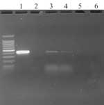
virus in oropharyngeal swab specimens from two chickens on day 1 PI .
Real-time RT-PCR results were confirmed by standard RT-PCR targeting the
same gene (primers: SARS clone 1b For 5´- TgACAgAgCCATgCCT-3´,
SARS clone 1b Rev 5´CAACggCATCATCAgA-3´) (Figure)
and sequencing of the amplified product. No infectious virus was isolated
from any of the birds at any time from oropharyngeal or cloacal swab specimens,
plasma, or tissues. Histologic examination did not identify any specific
lesions. No anti–SARS-CoV–specific antibodies were detected in birds at
0 or 10 days after injection. Levels of SARS-CoV were detected corresponding
to the inoculated titers in chicken and turkey embryonating eggs by real-time
RT-PCR, but not by virus isolation.
These findings suggest that poultry were unlikely to have been infected
during the recent SARS-CoV outbreak and were unlikely to have played any
role as amplifiers in the animal markets of southern China. The low level
of virus detected by real-time RT-PCR from the chickens and the failure
to isolate virus from embryonating chicken and turkey eggs suggest that
the detected virus was residual inoculum or nonviable virus and that substantial
virus replication in the poultry was unlikely. In addition, this SARS-CoV
was of low tissue culture passage, i.e., third passage in Vero E6 cell,
which minimized the potential for increased cell culture adaptation and
concomitant decrease in vivo replication. Using the original or second
tissue culture passage would unlikely have resulted in substantial replication
in poultry. However, the virus used in these experiments, the Urbani SARS-CoV,
had a 29-nt deletion in the genome. Whether the GZ01 human virus or those
from civet cats and raccoon dog containing the extra 29 nt would infect
and amplify in poultry would be of interest for future research.
Acknowledgments
We thank Suzanne
DeBlois and Scott Lee for excellent technical assistance.
Funding for this
study was provided by the U.S. Department of Agriculture, Agricultural
Research Service CRIS project #6612-32000-039.
Dr. Swayne is a
veterinary pathologist and the director of the Southeast Poultry Research
Laboratory of the Agricultural Research Service, U.S. Department of
Agriculture. His research focuses on pathobiology and control of exotic
and emerging viral diseases of poultry and other birds, principally
highly pathogenic avian influenza, avian metapneumovirus, and West Nile
virus.
References
- World Health Organization. Cumulative number of reported
probable cases of SARS. [accessed July 6, 2003]. Available from: http://www.who.int/csr/sars/country/2003_07_11/en/
- Peiris JS, Lai ST, Poon LL, Guan Y, Yam LY, Lim W, et al. Coronavirus
as a possible cause of severe acute respiratory syndrome. Lancet
2003;361:1319–25.
- Ksiazek TG, Erdman D, Goldsmith CS, Zaki SR, Peret T, Emery S, et
al. A
novel coronavirus associated with severe acute respiratory syndrome.
N Engl J Med 2003;348:1953–66.
- Drosten C, Gunther S, Preiser W, van der Werf S, Brodt HR, Becker
S, et al. Identification
of a novel coronavirus in patients with severe acute respiratory syndrome.
N Engl J Med 2003;348:1967–76.
- Fouchier RA, Kuiken T, Schutten M, van Amerongen G, van Doornum GJ,
van den Hoogen BG, et al. Aetiology:
Koch’s postulates fulfilled for SARS virus. Nature 2003;423:240.
- Field H. The role of animals in transmission of SARS. [accessed June
20, 2003]. Available from: http://www.who.int/csr/sars/conference/june_2003/materials/presentations/en/
- Guan Y, Zheng BJ, He YQ, Liu XL, Zhuang ZX, Cheung CL, et al. Isolation
and characterization of viruses related to the SARS coronavirus from
animals in southern China. Science 2003;302:276–8.
- Normile D, Enserink M. SARS
in China. Tracking the roots of a killer. Science 2003;301:297–9.
- Holmes KV. Coronaviruses. In: Granoff A, Webster RG, editors. Encyclopedia
of virology. San Diego: Academic Press; 1999. p. 291–8.
- Marra MA, Jones SJ, Astell CR, Holt RA, Brooks-Wilson A, Butterfield
YS, et al. The
genome sequence of the SARS-associated coronavirus. Science 2003;300:1399–404.
- Rota PA, Oberste MS, Monroe SS, Nix WA, Campagnoli
R, Icenogle JP, et al. Characterization
of a novel coronavirus associated with severe acute respiratory syndrome.
Science 2003;300:1394–9.
- Centers for Disease Control and Prevention. Influenza A (H9N2) infections
in Hong Kong. [accessed June 20, 2003]. Available from: http://www.cdc.gov/ncidod/diseases/flu/H9N2Info.htm
- World Health Organization. WHO news: Avian influenza virus reappears
in Hong Kong Special Administrative Region. Bull World Health Organ
2003;81:232.
- Centers for Disease Control and Prevention. Isolation
of avian influenza A (H5N1) from humans—Hong Kong, May–December, 1997.
MMWR Morb Mortal Wkly Rep 1997;46:1204–7.
- Barbeito MS, Abraham G, Best M, Cairns P, Langevin P, Sterritt WG,
et al. Recommended biocontainment features for research and diagnostic
facilities where animal pathogens are used. Rev Sci Tech Off Int Epiz
1995;14:873–87.
- Emery SL, Erdman DD, Meyer RF, Bowen MD, Tong S, Cook B, et al. Real-time
reverse transcription-polymerase chain reaction assay for the SARS-associated
coronavirus. Emerg Infect Dis 2004;10:311–6.
|