 |
| |
||
| |
|||||||||||||||||
|
|||||||||||||||||
|
|||||||||||||||||
|
EID Home | Ahead of Print | Past Issues | EID Search | Contact Us | Announcements | Suggested Citation | Submit Manuscript
|
|
Letter Rickettsia massiliae Human IsolationGiustina Vitale,* Serafino Mansueto,* Jean-Marc Rolain,† and Didier
Raoult† Suggested citation for this article To the Editor: The number of new rickettsial species that cause diseases in humans is rapidly increasing (1). Moreover, many of the species first described in ticks have been recently shown to be pathogenic. Of the 10 species or subspecies found to be pathogens after 1984, a total of 7 were first isolated from ticks (2). We report the first isolation of Rickettsia massiliae from a patient. The bacterium was isolated in Sicily in 1985 and identified in 2005.
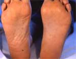
A 45-year-old man was hospitalized in Palermo, Italy, on June 6, 1985, for fever and a rash. He had been febrile since May 25 and did not respond to antimicrobial drug treatment using cefamezin, a first-generation cephalosporin. On examination, he had a necrotic eschar on his right ankle, a maculopapular rash on his palms and soles (Appendix Figure 1), and slight hepatomegaly. Leukocyte count was normal; he received tetracyclines for 13 days and fully recovered. He seroconverted (from 0 to 1:80 between day 11 and day 24) by indirect immunofluorescence to Rickettsia conorii (R. conorii spot, bioMérieux, Marcy l'Étoile, France). Four milliliters of heparinized blood sampled before treatment were inoculated in a 25-cm2 flask containing Vero cells and incubated at 33°C in a CO2 incubator (1). Direct immunofluorescence test on a sample of the patient's serum was positive 7 days later. The strain was stored for 20 years and tested in 2005 at the Unité des Rickettsies for identification, and R. massiliae was identified. DNA was extracted from the cell culture supernatant and used as template in 2 previously described polymerase chain reaction (PCR) assays that targeted a portion of the rickettsial ompA gene as well as a portion of the rickettsial gltA gene (3,4). Amplification products of the expected size were obtained from this extract but from no concurrently processed control materials, including 3 negative controls. DNA sequencing of the positive PCR products gave 100% identity with R. massiliae for ompA (GenBank accession no. RBU43792) and 99.9% homology for gltA (GenBank accession no. RSU 59720). R. massiliae was first isolated from Rhipicephalus ticks in Marseilles (5). It is transmitted transovarially in Rhipicephalus turanicus (2). R. massiliae is commonly found in Rhipicephalus sanguineus or R. turanicus in France, Greece, Spain (identified as Bar 29) (6), Portugal, Switzerland, Sicily (D. Raoult, unpub. data), Central Africa, and Mali (2). R. massiliae may be commonly associated with these ticks, which are distributed worldwide. R. massiliae is grouped phylogenically with Rickettsia rhipicephali and Rickettsia aeschlimannii (Appendix Figure 2). Bacteria from this group have a natural resistance to rifampin that is associated with an rpoB sequence that is different from that of other rickettsiae. This isolate was not tested for antimicrobial drug susceptibly (7). Rifampin resistance leads us to believe that this isolate may cause a Mediterranean spotted fever–like disease that was described in children in Spain (7,8). Serologic findings were recently reported that showed some patients in Barcelona, Spain, with reactions that indicate R. massiliae (B29 strain) rather than R. conorii (6). However, serologic reactions are only presumptive; isolation from a patient is the required to initially describe a new disease (9). This Sicilian index case shows that R. massiliae is a human pathogen. It contraindicates using rifampin to treat Mediterranean spotted fever in areas where R. massiliae is endemic, as it cannot as yet be differentiated from R. conorii infection. R. massiliae is a new example of a strain identified in ticks for several years before its first isolation from a human patient (10). The longest delay was observed for Rickettsia parkeri, which was isolated from ticks in 1939 but not from a patient until 2004. Many authors labeled R. parkeri a nonpathogenic rickettsia during this time (1). In the present case, the human isolate was obtained before the tick isolate but was not further identified. When this strain was isolated, R. conorii was the sole Rickettsia sp. found in ticks in southern Europe. Moreover, only 1 tickborne pathogenic Rickettsia sp. was believed to circulate in a single area. Since that time, several tickborne rickettsial diseases have been shown to exist in the same area, which prompted us to retrospectively identify this strain. The patient was reexamined in May 2005, after this identification. He is healthy and has no remaining antibodies against Rickettsia spp. References
Suggested citation
for this article: |
||||||||||||||||||||||||||||||
|
|
||||||||||||
|
||||||||||||
|
|
|
EID Home | Top of Page | Ahead-of-Print | Past Issues | Suggested Citation | EID Search | Contact Us | Accessibility | Privacy Policy Notice | CDC Home | CDC Search | Health Topics A-Z |
||
|
This page
posted December 6, 2005 |
||
|
Emerging
Infectious Diseases Journal |
||