|
|
Letter
Linguatuliasis in Germany
Dennis Tappe,* Ralf Winzer,* Dietrich W.
Büttner,† Philipp Ströbel,*August Stich,‡ Hartwig Klinker,* and Matthias Frosch* Ralf Winzer,* Dietrich W.
Büttner,† Philipp Ströbel,*August Stich,‡ Hartwig Klinker,* and Matthias Frosch*
*University of Würzburg, Würzburg, Germany; †Bernhard-Nocht-Institute
for Tropical Medicine, Hamburg, Germany; and ‡Medical Mission Hospital,
Würzburg, Germany
Suggested citation for this article
To the Editor: Pentastomids
or tongue worms are a unique group of vermiform parasites, phylogenetically
related to arthropods (1). Of the many pentastomid species, only a few, including Linguatula serrata, infect humans. The adult parasites are long, flat, or
annulated and have 4 hooks surrounding a central mouth. Adult L. serrata inhabit the nasal passages and paranasal sinuses of wild and domestic canids, which
serve as definitive hosts. Infective eggs containing larvae are discharged into
the environment by nasopharyngeal secretions and are ingested by herbivores, the
natural intermediate hosts. Humans can become dead-end intermediate hosts;
visceral linguatuliasis then develops (2) if infective eggs are
ingested. The liver is the organ most often involved (3–5), but the lung
(4,6,7) or other organs (4,8) may be affected. Parasites may also
be found in lymph nodes. In the viscera, the primary 4-legged larva molts
several times and eventually forms the legless nymph. Lesions due to Linguatula may be confused with malignancy, particularly in the lung (6).
We describe a recent infection with L.
serrata in Germany in a patient who had pulmonary symptoms and in whom
malignancy was suspected. The patient was a 39-year-old man of Russian origin
who had been living in Germany since 1999. He was admitted to the hospital with
weight loss, night sweats, chest pain, and coughing. He had been a smoker for
20 years, and his past medical history included pneumonia and sinusitis in 1989
during his military service at Lake Baikal, Russia. The patient had been living
in a farmhouse in Karaganda, Kazakhstan, until he immigrated to Germany.
A chest radiograph and computed tomographic
scan showed multiple, small lesions in both lungs. Malignancy was suspected,
and a bronchoscopy was performed. Numerous granulomatous nodules were
discovered. Thoracotomy was performed, and stringlike nodules on the pleural
surface were resected. Except for a mild eosinophilia (7%, 500 cells/μL), the leukocyte count
was normal. All other parameters, including C-reactive protein levels, angiotensin-converting enzyme, and tumor markers were normal. Histologic
examination of the nodules showed a targetoid appearance with a sharp demarcation
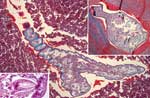
from the surrounding lung tissue by a thick fibrocollagenous capsule. In the
center of the nodules, a transverse section (Figure, right inset) and a
longitudinal section (Figure, main panel) of a parasite were visible. The
parasite had a chitinous cuticle ≈2.5 μm thick and cuticular spines 20–30 μm long. The spines and the
serrated aspect are characteristic for L. serrata, a pentastome. Ringlike
structures in the body wall were interpreted as sclerotized openings, a key
feature of pentastomes. In close contact to host tissue, a shed cuticle was visible
and assigned to the previous instar larva. The biometric data of the parasite were
comparable to those measured by others (6,9). Hooks, typical for the oral
armature of pentastomes, were found by serial sectioning (Figure, left inset). Except
for some subcuticular glands, the parasite's inner organs were no longer distinguishable.
The patient was initially treated with albendazole before the histologic
diagnosis of linguatuliasis was established. Findings from magnetic resonance
imaging of the abdomen were unremarkable, and no further lesions appeared
during 12 months of followup. Intermittent cough and chest pain remained,
possibly due to scar tissue and the remains of the nymphs.
At the beginning of the last century, visceral
linguatuliasis of humans occurred frequently in Germany. In 1904 and 1905,
among 400 autopsies in Berlin, 47 (11.8%) remains were infected with L. serrata (7). In contrast, reports of human infections are now rare. Our report
is the first recent case description in Germany. Where the patient acquired the
infection is unknown. L. serrata has a worldwide distribution. Recent
cases have been reported from China (4) and Italy (6). An
increasing number of infections can be suspected in the Western Hemisphere
because of incremental travel to linguatuliasis-endemic areas. Humans are
usually tolerant to nymphal pentastomid infections, and most patients are
asymptomatic (4). The living nymph provokes little inflammation, whereas
the death of the parasite leads to a prominent host response (2). Most
findings of visceral linguatuliasis are made at autopsy (4,6), and the
parasites are mainly located in the liver (3–5). Infection of the lung is
rare (6,7). The nymphs in human granulomas are typically degenerated at
the time of examination (3,6,9), but the cuticle with its associated
structures remains visible for some time (2). Histopathologic diagnosis
is guided by the presence of remnants of the cuticle with sclerotized openings and
by calcified hooks. Among pentastomids observed in humans, only L. serrata has prominent spines (2–4). In contrast to trematodes, the spines
protrude from the cuticle and do not end in the body wall of the parasite. Diagnosis
should be made etiopathologically, subetiopathologically, or presumptively on
the basis of whether entire nymphs, cuticle-associated structures, or pearly lesions
("Linguatula nodules" [10]) with targetoid appearance are found (4).
The differential diagnosis includes malignancies and tuberculosis because of the
radiologic coinlike appearance. On histologic examination, one must distinguish
between tissue-inhabiting diptera larvae, infections with metacestodes,
trematodes, tissue filariids, and gnathostomiasis. Once diagnosis is established,
no treatment is necessary (3) for the parasites will degenerate after
some time, and no effective antiparasitic therapy exists. Avoiding contact with
canine saliva and drinking water used by dogs or wild canids prevents this
infection.
References
- Lavrov DV, Brown WM, Boore JL. Phylogenetic position of the Pentastomida and (pan) crustacean relationships.
Proc Biol Sci. 2004;271:537–44.
- Baird JK, Carey JC. Pentastomiasis. In: Connor
DH, Chandler FW, editors. Pathology of infectious diseases. Stamford (CT):
Appleton & Lange; 1997. p. 1671–4.
- Baird JK, Kassebaum LJ, Ludwig
GK. Hepatic granuloma in a man from North America caused by a nymph
of Linguatula serrata. Pathology. 1988;20:198–9.
- Ma KC, Qiu MH, Rong YL. Pathological differentiation of suspected cases of pentastomiasis in China.
Trop Med Int Health. 2002;7:166–77.
- Gardiner CH, Dyke JW, Shirley SF. Hepatic granuloma due to a nymph of Linguatula serrata in a woman from
Michigan: a case report and review of the literature. Am J Trop Med Hyg.
1984;33:187–9.
- Pampiglione S, Gentile A, Maggi
P, Scattone A, Sollitto F. A nodular pulmonary lesion due to Linguatula
serrata in an HIV-positive man. Parassitologia. 2001;43:105–8.
- Koch M. Zur Kenntnis des Parasitismus der
Pentastomen. Verh Dtsch Ges Pathol. 1906;10:265–79.
- Lang Y, Garzozi H, Epstein Z,
Barkay S, Gold D, Lengy J. Intraocular pentastomiasis causing
unilateral glaucoma. Br J Ophthalmol. 1987;71:391–5.
- Lazo RF, Hidalgo E, Lazo JE,
Bermeo A, Llaguno M, Murillo J, et al. Ocular linguatuliasis in
Ecuador: case report and morphometric study of the larva of Linguatula
serrata. Am J Trop Med Hyg. 1999;60:405–9.
- St. Symmers WC, Valteris K. Two cases of human
infestation by larvae of Linguatula serrata. J Clin Pathol. 1950;3:212–9.
Suggested citation
for this article:
Tappe D, Winzer R, Büttner DW, Ströbel P, Stich A, Klinker H, et al. Linguatuliasis
in Germany [letter]. Emerg Infect Dis [serial on the Internet]. 2006 Jun [date
cited]. Available from http://www.cdc.gov/ncidod/EID/vol12no06/05-1413.htm
|