|
|
Dispatch
Rickettsia mongolotimonae
Infection in South Africa
Anne-Marié Pretorius* and Richard J. Birtles†
*University of the Free State, Bloemfontein, South Africa; and †University
of Liverpool, Liverpool, United Kingdom
Suggested citation
for this article:
Pretorius A-M, Birtles RJ. Rickettsia mongolotimonae infection
in South Africa. Emerg Infect Dis [serial online] 2004 Jan [date
cited]. Available from: URL: http://www.cdc.gov/ncidod/EID/vol10no1/02-0662.htm
We report the first
laboratory-confirmed case of Rickettsia mongolotimonae infection
in Africa. The patient sought treatment for an eschar on his toe; lymphangitis,
severe headaches, and fever subsequently developed. After a regimen
of doxycycline, symptoms rapidly resolved. R. mongolotimonae
infection was diagnosed retrospectively by serologic tests and molecular-based
detection of the organism in biopsy specimens of eschar material.
Rickettsioses are infections of emerging medical importance, particularly
in southern Africa, where an increasing number of cases are being encountered
among both residents and tourists (1). Three Rickettsia
species have been associated with human disease in South Africa to date.
Rickettsia conorii has long been recognized as the agent of Mediterranean
spotted fever, and more recently, a newly recognized species, R. africae,
has been identified as the agent of African tick-bite fever. In 2002,
the first case report of a patient infected with R. aeschlimannii
was published (2). In addition to these recognized pathogens,
Rickettsia species, including R. mongolotimonae, have been
detected in human-biting arthropods in Africa. This species (3)
was first encountered in Hyalomma asiaticum ticks in Inner Mongolia
in 1991 (4) but has subsequently been associated with
human infections in southern France (5) and, perhaps
of most relevance to this report, has been detected in H. truncatum
ticks collected from cattle in Niger (6). This species
of tick, which at least during its immature life stages parasitizes migratory
birds, is widely distributed in many African countries, including South
Africa (7).
The Study
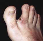
In September 2002, a 34-year-old (HIV-seronegative) construction worker,
working near Ellisras in South Africa’s Northern Province, discovered
a lesion on the inside of the second toe on his right foot (Figure);
subsequently, severe headaches and high fever developed. He was examined
at a local hospital and found to have lymphangitis extending pretibially
from the lesion; as a result of his other symptoms, he was treated for
blood poisoning with ceftriaxone sodium, 1,000 mg once daily. During the
next 3 days, the lesion at the bite site (noted by the examining physician)
remained very sore, and the patient’s right inguinal lymph node became
enlarged and very painful. The patient then decided to return to his hometown
and sought treatment from his general practitioner (on day 5 after discovery
of the lesion). On examination, the lesion and lymphangitis were clearly
visible on the patient’s toe, although cellulitis and edema were not observed.
His inguinal lymph node had swollen to 3 cm in diameter, and he was still
febrile (38.5°C). Blood samples were then obtained as well as a biopsy
specimen from the lesion. A regimen of doxycycline, 100 mg per day orally,
for 5 days was prescribed and 1 day’s dosage was administered. The next
day, the patient was afebrile, and the lymphangitis had completely resolved.
In the laboratory, a Giemsa stain of a smear prepared from the patient’s
blood showed activated lymphocytes. A complete blood count showed thrombocytosis
(632 x 103/mL), but all other hematologic parameters were within
the normal range. Biochemical findings showed elevated levels of alanine
transaminase (66 IU/L), blood urea nitrogen (7.2 g/L), and triglycerides
(2.2 mmol/L); and decreased levels of chloride (96.3 mmol/L) and albumin
(38g/L); all other tests yielded results within the normal range. Testing
of the patient’s serum with the Weil-Felix test demonstrated an antibody
titer (80) only against the OX2 Proteus antigen, giving presumptive
evidence of a rickettsial infection. As a result, antirickettsial microimmunofluorescence
testing was performed (8). The serum did not yield significant
immunoglobulin (Ig) M titers against R. conorii or R. africae
antigens, but IgG titers of 64 were found by using both antigens. DNA
was extracted from the eschar biopsy specimen by using the QIAamp Tissue
kit (QIAGEN GmbH, Hilden, Germany) according to the manufacturer's instructions.
This DNA extract was used as template in a previously described polymerase
chain reaction assay targeting a Rickettsia spp. rOmpA fragment
(9). An amplification product was obtained from this
extract but not from any concurrently processed control materials. The
amplification product was purified; the nucleic acid sequences of both
strands were then determined. The sequence obtained from these efforts
was found to share >99% similarity with the corresponding rOmpA
fragment of R. mongolotimonae.
Conclusions
The combination of clinical and laboratory data yielded strong evidence
that the case described here was an infection of R. mongolotimonae,
the first reported in southern Africa. A single eschar is also typical
of R. conorii infections, but these are characterized by rash (Mediterranean
spotted fever), which the current patient did not have. Although R.
africae infections manifest only rarely as a rash, they are typified
by multiple eschars (10). This case description is also
very similar to that relating to a French patient infected with R.
mongolotimonae, who had lymphangitis and inguinal lymphadenopathy.
The serologic findings indicate exposure to a spotted fever group rickettsia
rather than to a specific species within this group, but the near identity
of the rOmpA sequence obtained from the patient’s eschar to that
of R. mongolotimonae provides a clear indication that this species,
rather than other spotted fever group rickettsiae, was present at the
site of the tick bite. Although no tick was found in association with
the patient’s eschar, his infection may have been acquired from a H.
truncatum, as this species is abundant in the region of the bushveld
where the patient had been working and is known to feed on humans (11).
Dr. Pretorius is
a medical scientist and lecturer. at the National Health Laboratory
Services, Faculty of Health Sciences, University of the Free State,
Bloemfontein, South Africa. Her research interests include the clinical
and epidemiologic features of vector-borne diseases, especially rickettsiae,
ehrlichiae, and bartonellae.
Dr. Birtles is a
senior lecturer at the Centre for Comparative Infectious Diseases, Department
of Veterinary Pathology, Faculty of Veterinary Science, University of
Liverpool, Liverpool, UK. His research interests are epidemiology, ecology
and pathogenicity of bartonellae and other arthropod-borne bacteria,
particularly in natural reservoir–vector systems.
References
- Raoult D, Fournier P-E, Fenollar F, Jensenius M, Prioe
T, De Pina JJ, et al. Rickettsia
africae, a tick-borne pathogen of travellers to sub-saharan Africa.
N Engl J Med 2001;344:1504–10.
- Pretorius A-M, Birtles RJ. Rickettsia
aeschlimannii: a new pathogenic spotted fever group rickettsia,
South Africa. Emerg Infect Dis 2002;8:874.
- Raoult D, Brouqui P, Roux V. A
new spotted-fever-group rickettsiosis. Lancet 1996;348:412.
- Yu X, Fan M, Xu G, Liu Q, Raoult D. Genotypic
and antigenic identification of two new strains of spotted fever group
rickettsiae isolated from China. J Clin Microbiol 1993;31:83–8.
- Fournier P-E, Tissot-Dupont H, Gallais H, Raoult D. Rickettsia
mongolotimonae: a rare pathogen in France. Emerg Infect Dis
2000;6:290–2.
- Parola P, Inokuma H, Camicas J-L, Brouqui P, Raoult D. Detection
and identification of spotted fever group rickettsiae and ehrlichiae
in African ticks. Emerg Infect Dis 2001;7:1014–7.
- Walker J. A
review of the Ixodid ticks (Acari, Ixodidae) occurring in southern Africa.
Onderstepoort J Vet Res 1991;58:81–105.
- Teysseire N, Raoult D. Comparison
of Western immunoblotting and microimmunofluorescence for diagnosis
of Mediterranean spotted fever. J Clin Microbiol 1992;30:455–60.
- Roux V, Fournier P-E, Raoult D. Differentiation
of spotted fever group rickettsiae by sequencing and analysis of restriction
fragment length polymorphism of PCR amplified DNA of the gene encoding
the protein rOmpA. J Clin Microbiol 1996;34:2058–65.
- Roux V, Raoult D. Rickettsioses
as paradigms of new or emerging infectious diseases. Clin Microbiol
Rev 1997;10:694–719.
- Horak IG, Fourie LJ, Heyne H, Walker JB, Needham
GR. Ixodid ticks feeding on humans in South Africa: with notes on preferred
hosts, geographic distribution, seasonal occurrence and transmission
of pathogens. Exp Appl Acarol 2002;27:113–36.
|