Skip Standard Navigation Links
Centers for Disease Control and Prevention
 CDC Home Search Health Topics A-Z
peer-reviewed.gif (582 bytes)
eid_header.gif (2942 bytes)
 EID Home | Ahead of Print | Past Issues | EID Search | Contact Us | Announcements | Suggested Citation | Submit Manuscript

Volume 9, Number 9, September 2003

Microbiologic Characteristics, Serologic Responses, and Clinical Manifestations in Severe Acute Respiratory Syndrome, Taiwan

Po-Ren Hsueh, * Cheng-Hsiang Hsiao,* Shiou-Hwei Yeh,† Wei-Kung Wang,* Pei-Jer Chen,* Jin-Town Wang,* Shan-Chwen Chang,* Chuan-Liang Kao,* Pan-Chyr Yang,* and The SARS Research Group of National Taiwan University College of Medicine and National Taiwan University Hospital
*National Taiwan University Hospital, National Taiwan University College of Medicine, Taipei, Taiwan; and †National Health Research Institute, Taipei, Taiwan


Figure 1.
  Back to article
 

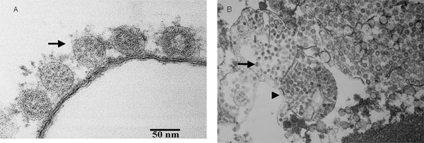
Figure 1. Thin-section electron micrograph of severe acute respiratory syndrome-associated coronavirus grown in Vero E6 cells. Panel A shows extracellular viral particles (arrow) lining the surface of the plasma membrane. Some spikes projecting from the envelope of the virus are seen. Panel B shows numerous spherical coronavirus particles (arrow) within dilated cytoplasmic vacuoles (arrowhead).


EID Home | Top of Page | Ahead-of-Print | Past Issues | Suggested Citation | EID Search | Contact Us | Accessibility | Privacy Policy Notice | CDC Home | CDC Search | Health Topics A-Z

This page last reviewed August 5, 2003

Emerging Infectious Diseases Journal
National Center for Infectious Diseases
Centers for Disease Control and Prevention