|
Dispatch
Outbreak of Neisseria
meningitidis, Edmonton, Alberta, Canada
Gregory J. Tyrrell,*† Linda Chui,* Marcia Johnson,‡ Nicholas
Chang,* Robert P. Rennie,*† James A. Talbot,*† and The Edmonton
Meningococcal Study Group[1]
*The Provincial Laboratory of Public Health for Alberta,
Alberta, Canada; †The University of Alberta, Edmonton, Alberta,
Canada; ‡The Capital Health Authority, Edmonton, Alberta, Canada
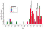
From December
1999 to April 2001, the greater Edmonton region had 61 cases of
invasive meningococcal infection, two fatal. The outbreak was
due to Neisseria meningitidis serogroup C, electrophoretic
type 15, serotype 2a. Analysis of the strains showed that 50 of
56 culture-confirmed cases were due to a single clone and close
relatives of this clone. This strain had not been previously identified
in the province of Alberta dating back to January 1997.
Neisseria meningitidis causes outbreaks of disease resulting
in severe illness and death. These outbreaks occur in persons in
their teens and early twenties; however, in some outbreaks, the
very young (<2 years of age) are also severely affected (1).
Persons >25 years of age appear to be less affected. North American
outbreaks are confined primarily to serogroup C strains and less
commonly to Y and W135 (2–5).
We report an outbreak of a serogroup C clone of N. meningitidis
in the Edmonton region of Alberta, Canada; the serogroup had a unique
restriction fragment length polymorphism (RFLP) pattern as determined
by pulsed-field gel electrophoresis (PFGE).
The
Outbreak
The Edmonton region has a mixed metropolitan and rural population
totaling 827,507 (6). From January 1997 to November
1999 (35 months), this region had 13 cases of culture-confirmed
invasive N. meningitidis disease (5 from blood, 6 from cerebrospinal
fluid [CSF], and 2 from joints) (Figure 1).
Serogroup determination, by the antiserum agar method previously
described, showed that these included two cases of serogroup B,
seven of serogroup C, two of serogroup W135, and two of serogroup
Y (7,8). During this period, the incidence of culture-confirmed
meningococcal disease did not exceed two cases per month (Figure
1). However, from December 1999 to April 2001, 61 cases of invasive
N. meningitidis disease occurred; 57 of these were confirmed
by culture and 4 on the basis of clinical findings, positive results
from an antigen detection assay, or both. The culture-confirmed
cases were from blood (51 cases) and CSF (6 cases). Of the 57 culture-confirmed
cases, 56 were serogroup C, and 1 was serogroup B (blood isolate).
In relation to clinical outcome, 43 (70.5%) of the 61 patients
fully recovered; 2 (3.3%) died (a 16-year-old woman and a 19-year-old
man, both infected with serogroup C) (Figure
1); 4 (6.6%) required amputations; 7 (11.5%) had severe scars;
and 9 (14.8%) had other sequelae such as knee pain, neurologic sequelae,
decreased hearing, decreased sensation at the extremities, and stiffness
in hands. The ages affected during the outbreak period ranged from
5 weeks to 77 years. Outbreak-associated patients were primarily
<24 years of age. Age breakdown showed that 10 (17.9%) of 56
confirmed serogroup C strains were in the birth- to 1-year age group
(Table). The conjugate vaccine for use in this
age group was licensed in Canada in May 2001 and was therefore not
available during the outbreak. The high number of cases in this
age group translates into an incidence rate of 50 per 100,000 (Table).
In comparison, the most recently published national data show the
rate for the group <1 year of age to be 12.9/100,000 for 1997
and 6.5/100,000 for 1998 (9). Also, age groups 15–19 and 20–24 showed
unusually high incidences of disease in this outbreak (Table).
Thirty patients with culture-confirmed disease were female, and
27 were male. Patients were scattered geographically throughout
the region, with no more than one case a close contact of another.
All contacts of patients were treated with rifampin. Except for
age group, no particular populations were determined to be at greater
risk for infection than other.
A vaccination campaign that targeted persons ages 2 to 19 was undertaken
in the region from February 14 to 28, 2000, using polysaccharide
quadravalent meningococcal vaccine; 168,000 children were immunized.
Because of a continuing higher-than-expected number of cases, the
vaccine was again offered in October 2000 (Figure
1). This time, the vaccine was offered to all previously unimmunized
2- to 24-year-olds (61,900 doses delivered in 6 days). In April
2001, vaccine was again offered to those 2-year-olds not previously
eligible in October 2000. Overall, 87% of people in the targeted
age group were vaccinated. After the vaccine campaigns, nine cases
of invasive meningococcal disease occurred in those eligible for
immunization but not immunized (total population of 2- to 24-year-olds
265,300). Nine cases also occurred in the immunized population,
for a calculated vaccine effectiveness of 84%.
Conclusions
Electrophoretic typing, serotyping, and serosubtyping performed
by the National Microbiology Laboratory, Population and Public Health
Branch of Health Canada, Winnipeg, Canada, showed that all serogroup
C strains belonged to electrophoretic type (ET)15, serotype 2a (10).
ET15 entered the Canadian population as early as 1986 in Ontario
and has since been demonstrated to be responsible for a number of
outbreaks in this country (9,11).
The most recent data for ETs in Canada date to 1997 and 1998 (9).
During 1997, ET15 accounted for 83.1% of strains analyzed. This
proportion increased in 1998 to 93.7%, indicating that ET is the
predominant type causing invasive disease in Canada. Serosubtyping
of serogroup C isolates in this outbreak showed variation in the
class 1 outer membrane protein (OMP1). Strains were either P1.2,5
(27 isolates), P1.2 (24 isolates), P1.15 (2 isolates), or P1.- (3
isolates). The two fatal cases were both P1.2,5.
Serogrouping, ET, and serosubtyping provided accurate characterization
of the circulating strains in the Edmonton region; however, they
failed to determine if the outbreak was clonal or if the increased
cases were due to unrelated N. meningitidis strains. To determine
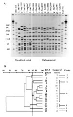
this, RFLP analysis via PFGE was used with minor modifications (12).
Bacterial cells were grown on sheep blood agar. The cells were scraped
off and placed in 10% formalin for 15 minutes for bacterial inactivation.
The cell number was standardized to an optical density of 1.4 at
A610. Lysozyme was added to 100 µL of cell suspension
at a final concentration of 0.2 mg/mL, mixed gently, and added to
1 mL of 1.6% (W/V) low-melting agarose. The mixture was then transferred
to the gel plug mold. The gel plug was suspended in Lysis II solution
(1 mg/mL lysozyme, 0.5% Brij 35, 0.2% sodium deoxycholate, and 0.5%
sodium lauryl sarcosine in Tris-EDTA buffer) for 1 hour at 37°C,
replaced with ESP buffer (0.25 M EDTA, pH 8.0, 1% sodium lauryl
sarcosine, 0.5 mg/mL Proteinase K), and incubated at 50°C for 2
hours. Slices of plug (1X5 mm) were digested by using 30 U of the
restriction endonuclease enzyme Spe I (GIBCO BRL, Burlington,
Ontario, Canada) for 2.0 hours. The restricted DNA was resolved
by PFGE with the following running conditions: initial switch time:
5.0 seconds, final switch time: 25 seconds at 6 V/cm with included
angle at 120°C for 20 hours. The gel was stained with ethidium bromide.
Analysis was performed by using the BioRad Gel Doc System (Bio-Rad
Laboratories, Mississauga, Ontario) and Molecular Analyst Software
(Bio-Rad). RFLP pattern numbers were assigned to each isolate with
one band difference in the RFLP profile.
RFLP analysis showed 15 distinguishable patterns (Figure
2A). Five of these RFLP patterns were seen only from January
1997 to November 1999 (patterns 6, 17, 20, 21, and 22). These patterns
were identified by retrospectively determining their RFLP profile
from archived strains. The remaining 10 were present only from December
1999 to April 2001 (patterns 3, 7, 8, 11, 12, 16, 25, 32, 34, and
44). Figure 2B shows a dendrogram analysis
of the 15 Spe I-generated RFLP profiles generated by using
a 1% tolerance. We used an 85% breakpoint to determine relatedness,
as reported by Popovic et al. (13). At the 85%
relatedness breakpoint, the RFLP profiles formed seven clusters.
RFLP patterns for the largest cluster (49 isolates-cluster 4) were
only seen during the outbreak period (December 1999 to April 2001).
Both deaths were associated with RFLP pattern 3 strains. Interestingly,
pattern 7 in cluster 3 was similar on visual examination to pattern
3 (Figure 2A). The first RFLP pattern (first
case) detected was pattern 3 (December 24, 1999) followed by pattern
7 (second case, December 29, 1999). Pattern 7 was not detected before
December 1999. These data suggest that patterns 3 and 7 arose concomitantly.
Even though these patterns appear close in time, pattern 3 and its
relatives resulted in 51 cases, whereas pattern 7 was isolated from
only 3 cases. Whether pattern 3 strains are more virulent than pattern
7 strains remains to be determined. We have also received reports
that this clone has caused disease in other regions of the province
of Alberta and in one other Western Canadian province in the same
period reported for our outbreak.
In conclusion, the Edmonton region in the province of Alberta,
Canada, had an outbreak of N. meningitidis caused by a clone
unique to this region. This clone was associated with increased
deaths and can readily spread beyond defined geographic boundaries.
Other provincial and state laboratories need to be able to recognize
this clone should it appear in their area.
Acknowledgments
We thank Jan Stoltz and Raymond Tsang for providing the electrophoretic
typing, subtyping, and serosubtyping analysis, and Susanna Schmink
for providing Neisseria meningitidis strain CDC 413.
Dr. Tyrrell is a clinical microbiologist in the Provincial Laboratory
for Public Health-Alberta. He holds academic appointments in the
Departments of Laboratory Medicine and Pathology and Medical Microbiology
and Immunology, University of Alberta. He is also the Director of
the National Centre for Streptococcus-Canada. His research interests
are streptococci and meningococci.
References
- Jackson LA, Schuchat A, Reeves MW, Wenger JD.
Serogroup
C meningococcal outbreaks in the United States, an emerging threat.
JAMA 1995;273:383–9.
- Schwartz B, Moore PS, Broome CV. The
global epidemiology of meningococcal disease. Clin Microbiol
Rev 1989;2:S118–S124.
- Tikhomirov E. Meningococcal meningitidis: global situation and
control measures. World Health Stat Q 40:98–108.
- Achtman M. Molecular epidemiology of epidemic meningtidis. Rev
Med Microbiol 1990;1:29–38.
- Apicella MA. Neisseria meningitidis. In: Mandel GL, Bennet
JE, Dolin R, editors. Principles and practice of infectious diseases.
5th ed. Philadelphia: Churchill Livingstone; 2000. p. 2228–41.
- Predy GN, Lightfoot P, Edwards J,Fraser-Lee N. How healthy are
we? Health status in the Capital Health Region-A technical report
2000. Edmonton, Alberta: Capital Health Authority; 2001.
- Ashton FE, Ryan A, Diena BB.
Improved antiserum agar for the serogroup differentiation of Neisseria
meningtiditis Y and w135. Can J Microbiol 1980;26:630–2.
- Craven DE, Frasch EE. Serogroup
identification of meningococci by a modified antiserum agar method.
J Clin Microbiol 1979;9:547–8.
- Squires SG, Pelletier L, Mungai M, Tsang R, Collins F, Stoltz
J. Invasive
meningococcal disease in Canada, 1 January 1997 to 31 December
1998. Can Commun Dis Rep 2000;26-21:177–82.
- Abdillahi H, Poolman JT. Whole-cell ELISA for typing Neisseria
meningitidis with monoclonal antibodies. FEMS Microbiol Lett
1987;48:367–71.
- Ashton FE, Ryan JA, Borczyk A, Caugant DA,
Mancino L, Huang D. Emergence
of a new virulent clone of Neisseria meningitidis serotype
2a that is associated with meningococcal group C disease in Canada.
J Clin Microbiol 1991;29:2489–93.
- Chang N, Chui L. A
standardized protocol for the rapid preparation of bacterial DNA
for pulsed-field electrophoresis. Diagn Microbiol Infect Dis
1998;31:275–9.
- Popvic T, Schmink S, Rosenstein NA, Ajello GW, Reeves MW, Plikaytis
B, et al. Evaluation
of pulsed-field gel electrophoresis in epidemiological investigations
of meningococcal disease outbreaks caused by Neisseria meningitidis
serogroup C. J Clin Microbiol 2001;39:75–85.
[1] The Provincial Laboratory of Public Health
for Alberta; Dora Lee, The Capital Health Authority; Kari Bergstrom,
Gerald Predy, Alberta Health and Wellness; Karen Grimsrud, Agnes
Honish and John Waters.
| Table.
Rates of Neisseria meningitidis disease in the Edmonton,
Canada, region (per 100,000)a |
|
| Rate |
Age (cases)
|
|
|
<1
|
2–4
|
5–9
|
10–14
|
15–19
|
20–24
|
25–34
|
35–59
|
>60
|
|
|
17-month periodb
|
50.0
(9)
|
9.7
(3)
|
5.3
(3)
|
6.8
(4)
|
35.7
(20)
|
10.6
(6)
|
4.0
(5)
|
2.3
(7)
|
1.7
(3)
|
|
Annualized
|
37.5
|
7.3
|
4.0
|
5.1
|
26.8
|
8.0
|
3.0
|
1.8
|
1.3
|
|
|
aPopulation=827,507 (6).
bDecember 1999 through April
2001
|
|