 |
| |
||
| |
|||||||||||||||||
|
|||||||||||||||||
|
|||||||||||||||||
|
EID Home | Ahead of Print | Past Issues | EID Search | Contact Us | Announcements | Suggested Citation | Submit Manuscript
|
|
Dispatch Intact pks15/1 in Non–W-Beijing Mycobacterium tuberculosis IsolatesAngkana Chaiprasert,*† Suggested citation for this article
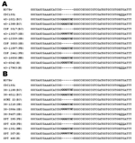
Two structurally related families of cell envelope lipids, phthiocerol diesters and phenolic glycolipids, are virulence factors of Mycobacterium tuberculosis and M. leprae. They are also produced by other slow-growing species, in particular the pathogenic species M. marinum, M. ulcerans, and members of M. tuberculosis complex (1). Phthiocerol diesters are composed of a mixture of long chain β-diols that are esterified by multimethyl-branched fatty acids. Depending on the asymmetric centers bearing the methyl branches (D or L series), the fatty acids are called mycocerosic or phthioceranic acids, respectively, and the corresponding complex lipids are named dimycocerosates of phthiocerol (DIMs) or diphthioceranates of phthiocerol (DIPs) (1). The phenolic glycolipids (PGLs) consist of a lipid core similar to those of DIMs or DIPs but ω-terminated by an aromatic nucleus that is glycosylated by type- or species-specific mono-, tri-, or tetrasaccharide. Several lines of evidences suggest that PGLs are involved in the pathogenesis of mycobacterial infections. PGL-1 from M. leprae inhibits the proliferation of T lymphocytes after stimulation with concanavalin A (2). Moreover, PGL-1 seems to be associated with resistance to intracellular killing by macrophages (3) and promotes phagocytosis of M. leprae by macrophages and Schwann cells by binding to complement component C3 or laminin α2 chain, respectively (4,5). Similarly, PGLs produced by a subset of M. tuberculosis isolates inhibit the host Th1-type T-cell and cytokine response (6). All M. tuberculosis strains tested that produce PGLs belong to the W-Beijing family and show a "hypervirulent" phenotype, in comparison with the clinical isolate M. tuberculosis CDC1551 and the laboratory strain M. tuberculosis H37Rv in the murine model (6) and rabbit model of meningitis (7). Previous study identified the involvement of the gene pks15/1 in the biosynthesis of PGLs; disruption of this gene generated a PGL-deficient mutant (8). Sequence alignment of the pks15/1 gene, when compared to the non–PGL-producing strains, M. tuberculosis H37Rv, Erdman, Mt103, and CDC1551, that contain 2 open reading frames [pks1 (Rv2946c) and pks15 (Rv2947c)], showed a 7-bp insertion in PGL-producing strains M. tuberculosis strain 210, belonging to the W-Beijing family, and M. canetti, whereas M. bovis and M. bovis BCG contained only a guanine insertion. This 7-bp or 1-bp insertion causes a frameshift mutation in the pks15, resulting in an intact pks15/1 with additional codons (8). Similar results have been shown in other W-Beijing strains, M. tuberculosis HN878, W4, and W10, which contain the 7-bp insertion and produce PGLs (6). In Thailand, the Beijing genotype is the predominant genotype among tuberculosis (TB) patients, particularly in patients with TB meningitis (unpub. data), which suggests recent transmission of this genotype in the country. Similarly, the Beijing genotype has been found frequently in Asia (9–11). Previous studies have shown that the M. tuberculosis strains belonging to this genotype contain an intact pks15/1 and can produce PGLs that associated with the hypervirulent phenotype (6,7). The goal of our study was to determine whether the hypervirulence of the W-Beijing strains due to the ability to produce PGLs is unique among this family by investigating the pks15/1 gene of the Beijing strains compared to other strains that can cause diseases similar to those caused by Beijing strains. The StudyOne hundred forty-seven clinical isolates of M. tuberculosis were obtained from the Molecular Mycobacteriology Laboratory, Department of Microbiology, Faculty of Medicine, Siriraj Hospital, Mahidol University, Thailand, and the T-2 project from 1997 to 2001 (Table). These strains were isolated from 74 cerebrospinal fluid (CSF) samples and 73 sputum samples from 147 different patients. DNA from these isolates was isolated by an enzymatic method and submitted for genotyping by performing the IS6110 restriction fragment length polymorphism with the standard method (12) and for sequencing the pks15/1 region (8).
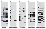
Using the genotyping results, we categorized M. tuberculosis isolates into Beijing, single-banded, few-banded (2–5 bands), Nonthaburi, and heterogeneous with >5 bands (Table and Figure 1), as recently reported (13,14). All M. tuberculosis genotypes were sequenced around the junction of pks15 and pks1 (corresponding to the M. tuberculosis H37Rv sequence) to determine whether they contained an intact pks15/1 or separated pks15 and pks1. Unexpectedly, the results showed that the 7-bp insertion of pks15 that causes a frameshift mutation resulting in an intact pks15/1 was found in most strains of all genotypes, except the heterogeneous group with >5 bands (Table and Figure 2). ConclusionsThe intact pks15/1 has been shown to be responsible for the production of phenolic glycolipids and is seemingly found in M. tuberculosis W-Beijing family, but it was not found in M. tuberculosis CDC1551 and H37Rv (8). Previous studies suggested that PGLs produced by the M. tuberculosis W-Beijing family were associated with the hypervirulent phenotype by inhibiting the innate immune response (6,7). The intact pks15/1 has also been shown to be nonpolymorphic in the W-Beijing family; it was found in all 102 W-Beijing strains tested (15). From this observation, we hypothesized that if the ability to produce PGLs is among the factors that make this family more virulent than others, the intact pks15/1 should be absent in strains other than the W-Beijing family. Our results showed that the 7-bp insertion of the pks15/1 was not only present in the W-Beijing family but also in other M. tuberculosis genotypes. Although almost all Beijing strains contain the intact pks15/1 (≈97%), 38.5%–100% of strains of other genotypes also contain it. These strains could, therefore, produce PGLs and cause both pulmonary and disseminated diseases as the W-Beijing strains do. Our results showed no significant difference in the percentage of M. tuberculosis isolates with an intact pks15/1 gene between CSF isolates (65 [87.8%] of 74) and sputum isolates (62 [84.9%] of 73). The hypothesis that the hypervirulence of the W-Beijing family is solely attributable to pks15/1 is still inconclusive. This family may have only recently been transmitted globally and may have had more chances to cause infections and disease than other families. Although PGLs are involved in the hypervirulence of the PGL-producing strains, they are not a unique characteristic of the W-Beijing family. If W-Beijing strains are more virulent than others, other virulence determinants besides PGLs must be responsible for the hypervirulent phenotype. Acknowledgments
References
Suggested citation
for this article: |
||||||||||||||||||||||||||||||||||||||||
|
|
||||||||||||
|
||||||||||||
|
|
|
EID Home | Top of Page | Ahead-of-Print | Past Issues | Suggested Citation | EID Search | Contact Us | Accessibility | Privacy Policy Notice | CDC Home | CDC Search | Health Topics A-Z |
||
|
This page
posted April 11, 2006 |
||
|
Emerging
Infectious Diseases Journal |
||