ISSN: 1080-6059
Volume 14, Number 8–August 2008
Dispatch
Genotyping Rickettsia prowazekii Isolates
Yong Zhu,* Aaron Medina-Sanchez,* Donald Bouyer,* David H. Walker,* and Xue-jie Yu* ![]()
*University of Texas Medical Branch, Galveston, Texas, USA
Suggested citation for this article
Abstract
We developed a typing method that can differentiate 8 strains of Rickettsia prowazekii into 7 genotypes.
This method can be used to type and trace the origin of R. prowazekii isolated from samples collected during epidemics
after a bioterrorism attack.
Rickettsia prowazekii is the causative agent of epidemic typhus and also a potential bioterrorism agent. The disease may occur in epidemics when social, economic, or political systems are disrupted and expose a large population such as refugees to louse infestation due to lack of hygiene. Recent outbreaks of typhus have occurred in Burundi, Algeria, Peru, and Russia (1,2). R. prowazekii is transmitted by the human body louse, Pediculus humanus corporis, in the human cycle. Sylvatic typhus associated with R. prowazekii has been documented in the eastern United States. However, it is not clear whether R. prowazekii transmission to humans from flying squirrels results from the bite of fleas or lice or contaminated arthropod fecal material (3,4). Reemergence of epidemic typhus and the potential use of R. prowazekii in bioterrorist attacks requires a molecular method that can type isolates and trace the origin or epidemiology of the disease.
The Study
Our objective was to identify a minimal gene set in which PCR amplification and sequencing would allow the efficient differentiation of R. prowazekii strains for diagnostic purposes. Using BLAST analysis (www.ncbi.plm.nih.gov/blast/b12seq/wblast2.cgi) to identify target DNA sequences for genotyping, we compared the genomic sequences of Madrid E strain (E strain, NC_000963) (5) with those of Nuevo Leon strain, a new tick isolate of R. prowazekii (6), which was sequenced recently (unpub. data). We identified 6 loci with insertion or deletion in 1 of 2 strains. PCR primers were designed from the target sequences and used to amplify DNA from 8 strains of R. prowazekii, including human isolates Addis Ababa, Breinl, Cairo, and E strain; a guinea pig isolate of Evir strain (7); a tick isolate (ZRS) from Ethiopia (8); and 2 flying squirrel isolates (GvV-250 from Virginia and GvF-16 from Florida) (Table 1) (4). Rickettsial genomic DNA was extracted from the R. prowazekii–infected L929 cells or infected yolk sacs of embryonated chicken eggs by using the GenElute Mammalian Genomic DNA Miniprep kit (Sigma-Aldrich, St. Louis, MO, USA) according to the manufacturer's instructions.
For designing the primers (Table 1), we used Primer 3.0 software (http://frodo.wi.mit.edu/cgi-bin/primer3/primer3_www.cgi); primers were synthesized. Two microliters of the DNA preparation were amplified in a 50-μL RED taq ReadyMIX PCR reaction (Sigma-Aldrich). The following conditions were used for amplification: an initial 5 min of denaturation at 94°C followed by 30 cycles of denaturation for 30 s at 94°C, annealing for 30 s at 53 °C, and extension for 1 min at 72°C. Amplification was completed by holding the reaction mixture for 2 min at 72°C. PCR products were directly sequenced with PCR primers for both strands. PCR amplification and DNA sequencing were performed twice for each gene of each R. prowazekii strain. A PCR reaction without template DNA was included as a negative control in each PCR.
DNA sequences were aligned by using DNASTAR Lasergene software, version 6.0 (DNASTAR, Inc., Madison, WI, USA). The sequences amplified by 6 pairs of primers from each strain were joined together to form a concatenated sequence for each strain. A multiple alignment of the concatenated sequences was constructed by using ClustalW (www.ebi.ac.uk/clustalw) and was analyzed by using the neighbor-joining method in PAUP 4.0 Beta (Sinauer Associate, Inc., Sunderland, MA, USA). Bootstrap was estimated for neighbor-joining trees by 1,000 resamplings. The sequences reported here were assigned consecutive GenBank accession numbers from EU192931 to EU192949.
Conclusions
We amplified the 6 loci from all 8 R. prowazekii strains and compared the corresponding sequences of each strain to identify the variations among strains. Three loci were intergenic spacers (rp272/rp273, rp308/rp309, and rp691/rp692), and 2 loci were pseudogenes (rp181 and rp195) in all R. prowazekii strains. We also sequenced rp028, the methyltransferase gene, because we wanted to know if this gene was inactivated in any virulent strain of R. prowazekii. Pseudogene rp028 was inactivated in a virulent E strain but not in its virulent revertant Evir strain (9). Coincident with inactivation of the methyltransferase gene, E strain is deficient in methylation of surface proteins (10,11).
Our result shows that a single nucleotide insertion at position 732 in rp028 occurred only in E strain among the tested R. prowazekii strains (Table 2). However, single nucleotide polymorphism (SNP) existed in rp028 among strains of R. prowazekii and was very useful in the differentiation of R. prowazekii strains (Table 2). Apparently none of these nucleotide substitutions caused attenuation of E strain because the E strain and Evir strain were identical at these sites.
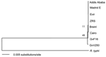 |
Figure. Phylogenic tree of Rickettsia prowazekii strains generated by using the concatenated sequences of 6 loci from each strain. R. typhi sequences were used to root the tree. |
DNA sequence comparison and phylogenetic analysis of the concatenated sequences indicated that the R. prowazekii strains were grouped together by geographic location and source of isolation (Table 2, Figure). Two flying squirrel isolates from the United States were differentiated by a single nucleotide substitution at position 480 in rp028. E strain and its revertant Evir strain differed by a single nucleotide insertion in E strain at position 732 in rp028, which we reported previously (9). Breinl and Cairo strains were closely related but were differentiated by several deletion/insertion mutations in rp181 and the spacer between rp272 and rp273. The cattle tick isolate ZRS and the human isolate Addis Ababa, both from Ethiopia, were identical in all 6 loci. ZRS strain and Addis Ababa strain were phylogenetically more closely related to E/Evir strains than other strains (Figure). There was only a single nucleotide difference between ZRS/Addis Ababa strains and Evir strain (Table 2).
Genotyping of R. prowazekii has been explored recently. Zhu et al., using intergenic spacers rpmE/tRNAfMet and serS/virB4, differentiated 5 strains and PCR amplicons from 10 body lice of R. prowazekii into 4 genotypes (12). Ge et al. showed that R. prowazekii Breinl strain and E strain were different in the rp084 gene, which was deleted from the Breinl strain (13). However, using the rpmE/tRNAfMet intergenic spacer, we were able to classify the 8 strains of R. prowazekii tested into only 2 genotypes. Genotype 1 contains Breinl strain and genotype 2 includes all other strains. All 8 strains were identical in the serS/virB4 spacer. With the exception of R. prowazekii Breinl strain, rp084 was not deleted from any strains of R. prowazekii tested in our study. Conversely, using our methods, the 8 strains of R. prowazekii can be differentiated into 7 genotypes. ZRS and Addis Ababa strains are the only isolates that cannot be differentiated with our method. Because all R. prowazekii ZRS and Addis Ababa strains originated from Ethiopia, it is reasonable to believe that they might be genetically identical. Ge et al. recently showed that 5 R. prowazekii strains, including Breinl, Cairo, E, GvV257, and GvF12 were different from each other by 1 to 4 SNPs in ompB and sca4, respectively (14). However, the differentiation of R. prowazekii based on SNPs between closely related strains may be complicated by PCR and sequence errors. Conversely, our method confers more confidence in the validation of the mutations because we differentiated all strains except for 2 flying squirrel strains by insertion and deletion mutations, which are rarely generated by PCR or sequence errors.
Our method provides a technique for typing and tracing the origin of new R. prowazekii isolates. This method will have a broad use in the biodefense against and the molecular epidemiology of R. prowazekii and in detection of laboratory cross-contamination of R. prowazekii strains.
Acknowledgment
We are grateful to Dr Zhikai Zhang for help in DNA sequencing.
This study was supported by a grant (U01AI71283) from the National Institute of Allergy and Infectious Diseases.
Dr Zhu is a postdoctoral fellow in the Department of Pathology, University of Texas Medical Branch at Galveston. His research interests are in the genotyping and molcecular biology of Rickettsia spp.
References
- Raoult D, Woodward T, Dumler JS. The history of epidemic typhus. Infect Dis Clin North Am. 2004;18:127–40. PubMed DOI
- Raoult D, Roux V, Ndihokubwayo JB, Bise G, Baudon D, Marte G, et al. Jail fever (epidemic typhus) outbreak in Burundi. Emerg Infect Dis. 1997;3:357–60.
- Bozeman FM, Sonenshine DE, Williams MS, Chadwick DP, Lauer DM, Elisberg BL. Experimental infection of ectoparasitic arthropods with Rickettsia prowazekii (GvF-16 strain) and transmission to flying squirrels. Am J Trop Med Hyg. 1981;30:253–63.
- Duma RJ, Sonenshine DE, Bozeman FM, Veazey JM Jr, Elisberg BL, Chadwick DP, et al. Epidemic typhus in the United States associated with flying squirrels. JAMA. 1981;245:2318–23. PubMed DOI
- Andersson SG, Zomorodipour A, Andersson JO, Sicheritz-Ponten T, Alsmark UC, Podowski RM, et al. The genome sequence of Rickettsia prowazekii and the origin of mitochondria. Nature. 1998;396:133–40. PubMed DOI
- Medina-Sanchez A, Bouyer DH, Cantara-Rodriguez V, Mafra C, Zavala-Castro J, Whitworth T, et al. Detection of a typhus group Rickettsia in Amblyomma ticks in the state of Nuevo Leon, Mexico. Ann N Y Acad Sci. 2005;1063:327–32. PubMed DOI
- Balayera NM, Nikolskaya VN. Enhanced virulence of the vaccine strain E of Rickettsia prowazekii on passaging in white mice and guinea pigs. Acta Virol. 1972;16:80–2.
- Reiss-Gutfreund RJ. The isolation of Rickettsia prowazeki and mooseri from unusual sources. Am J Trop Med Hyg. 1966;15:943–9. PubMed
- Zhang JZ, Hao JF, Walker DH, Yu XJ. A mutation inactivating the methyltransferase gene in avirulent Madrid E strain of Rickettsia prowazekii reverted to wild type in the virulent revertant strain. Vaccine. 2006;24:2317–23. PubMed DOI
- Ching WM, Carl M, Dasch GA. Mapping of monoclonal antibody binding sites on CNBr fragments of the S-layer protein antigens of Rickettsia typhi and Rickettsia prowazekii. Mol Immunol. 1992;29:95–105. PubMed DOI
- Ching WM, Wang H, Davis J, Dasch GA. Amino acid analysis and multiple methylation of lysine residues in the surface protein antigen of Rickettsia prowazekii. In: Angeletti RH, editor, Techniques in protein chemistry, Vol. IV. San Diego: Academic Press. 1993:307–14.
- Zhu Y, Fournier PE, Ogata H, Raoult D. Multispacer typing of Rickettsia prowazekii enabling epidemiological studies of epidemic typhus. J Clin Microbiol. 2005;43:4708–12. PubMed DOI
- Ge H, Chuang YY, Zhao S, Tong M, Tsai MH, Temenak JJ, et al. Comparative genomics of Rickettsia prowazekii Madrid E and Breinl strains. J Bacteriol. 2004;186:556–65. PubMed DOI
- Ge H, Tong M, Jiang J, Dasch GA, Richards AL. Genotypic comparison of five isolates of Rickettsia prowazekii by multilocus sequence typing. FEMS Microbiol Lett. 2007;271:112–7. PubMed DOI
Figure
Tables
Table 1. Primers for 6 loci of Rickettsia
prowazekii genomic DNA sequences
Table 2. Genotypes of Rickettsia
prowazekii strains determined by nucleotide mutation in multiple loci
Suggested Citation for this Article
Zhu Y, Medina-Sanchez A, Bouyer D, Walker DH, Yu X. Genotyping Rickettsia prowazekii isolates. Emerg Infect Dis [serial on the Internet]. 2008 Aug [date cited]. Available from http://www.cdc.gov/EID/content/14/8/1300.htm
DOI: 10.3201/eid1408.080444
Please use the form below to submit correspondence to the authors or contact them at the following address:
Xue-jie Yu, Unviersity of Texas Medical Branch, 301 University Blvd, Galveston, TX 77555–0609, USA; email: xuyu@utmb.edu
Please contact the EID Editors at eideditor@cdc.gov
The opinions expressed by authors contributing to this journal do not necessarily reflect the opinions of the U.S. Department of Health and Human Services, the Public Health Service, the Centers for Disease Control and Prevention, or the authors' affiliated institutions. Use of trade names is for identification only and does not imply endorsement by any of the groups named above.
This page posted July 25, 2008
Centers for Disease Control and Prevention, 1600 Clifton Rd, Atlanta, GA 30333, U.S.A
Tel: (404) 639-3311 / Public Inquiries: (404) 639-3534 / (800) 311-3435