2007
News Archive 2008
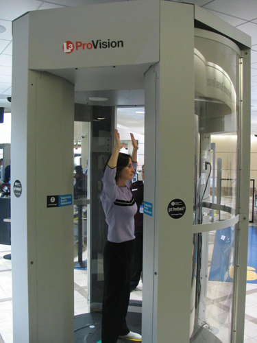
Caption: ProVision™, developed at PNNL. (Click image to download hi-res version).
Across the United States, passengers are entering a space age-looking machine that can detect objects hidden on their bodies.
The system, known as ProVision™, was developed at Pacific Northwest National Laboratory (PNNL) and is now in 10 airports; by 2009, the Transportation Security Administration (TSA) will install a total of 120 more systems in 21 airports.
"Using the millimeter wave technology in airports is one of the first significant changes we've made to checkpoint technology since the 1970s," said Ellen Howe, TSA's assistant administrator for strategic communications and public affairs. "The technology is being more broadly deployed in the airport environment because of its ability to detect materials that cannot be found by the walk thru metal detector."
PNNL's millimeter wave technology was just a gleam in researcher Doug McMakin's eye in the 1980s.
By the early 1990s, he and a team developed a prototype that was tested at Seattle-Tacoma International Airport and demonstrated for companies in the U.S., Japan, Canada, and Israel.
It wasn't until 2002, however, that the technology actually moved toward the marketplace.
Battelle, which operates PNNL for the Department of Energy, licensed the patented technology to SafeView, Inc., a startup company formed in 2002 to commercialize the scanning technology into noninvasive security "portals."
In 2006, L-3 Communications acquired SafeView, Inc., and started selling the ProVision systems to TSA for use in airport screening.
By the end of 2008, 21 U.S. airports will have ProVision systems, and TSA will purchase and install 80 more in 2009.
"You can see the ear-to-ear grin on my scanned image," said Doug McMakin, one of the technology developers. "It's very gratifying to see your work in the marketplace, particularly when it's being used in public places and for important purposes like airport security."
The millimeter wave technology uses non-harmful, ultrahigh-frequency radio waves to penetrate clothing; the reflection off the body allows system operators to see hidden objects—even nonmetallic ones, which is just one of the advantages of the system.
Other advantages are its scanning speed (about 30 seconds) and the fact that it doesn't require person-to-person contact.
"I'd certainly prefer to be scanned rather than patted down," said McMakin.
-This article was provided by Pacific Northwest National Laboratory.-Undertaken in collaboration with the University of California, Davis Cancer Center, the compact proton therapy system would fit in any major cancer center and cost about one-fifth as much as a full-scale machine. This innovation has garnered researchers at Lawrence Livermore National Laboratory an award for excellence in technology transfer by the Federal Laboratory Consortium in 2008.
Lawrence Livermore National Laboratory (LLNL) licensed the technology in February 2007 to TomoTherapy Incorporated (NASDAQ: TTPY) of Madison, Wis., through an agreement with the Regents of the University of California.
Proton therapy is considered the most advanced form of radiation therapy available, but size and cost have limited the technology’s use to only six centers in the United States and 25 worldwide. Together, they have treated an estimated 40,000 patients.
TomoTherapy will fund development of the first clinical prototype, which will be tested on patients at the UC Davis Cancer Center. If clinical testing is successful, TomoTherapy will bring the machines to market.
Conventional proton therapy facilities use cyclotrons and synchrotrons to deliver protons, and these large facilities can require 90,000 square feet and cost more than $100 million to build.
The Livermore team has overcome the size obstacle by using a dielectric wall accelerator technology that enables proton particles to be accelerated to an energy of at least 200 million electron volts within a lightweight, insulator-based structure about 6.5 feet long.
LLNL’s dielectric wall accelerator advance grew out of work to develop compact, high-current accelerators, such as flash X-ray radiography sources for nuclear weapons stockpile stewardship.
The current patent portfolio consists of 20 inventions. More inventions are expected as full-scale feasibility is demonstrated.
For more information, visit the Industrial Partnerships Office and Lawrence Livermore National Laboratory.
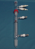
-This article was provided by Lawrence Livermore National Laboratory.-The Savannah River National Laboratory's Glass Development Laboratory, which designs and fabricates unique scientific glass apparatus, has a long history of helping researchers develop more effective tools for experimental work. Among their innovations is the newly patented Two Part Condenser for Varying the Rate of Condensing, invented by Gary Dobos of the Glass Development Lab. This condenser design enables the unit to effect different rates of condensing during experiments. It was designed as a way to increase condensing rates as experiments were in progress. The design offers a second condenser that can be inserted into the primary condenser to increase efficiency without disassembling the original set-up or interrupting the test.
-This article was provided by Savannah River National Laboratory.-First step toward three-dimensional catalytic, magnetic, and/or optical nanomaterials
Caption: Researchers Matthew Maye, Niels van der Lelie, Oleg Gang, and Dmytro Nykypanchuk. (Click image to download hi-res version).UPTON, NY - In an achievement some see as the "holy grail" of nanoscience, researchers at the U.S. Department of Energy's Brookhaven National Laboratory have for the first time used DNA to guide the creation of three-dimensional, ordered, crystalline structures of nanoparticles (particles with dimensions measured in billionths of a meter). The ability to engineer such 3-D structures is essential to producing functional materials that take advantage of the unique properties that may exist at the nanoscale - for example, enhanced magnetism, improved catalytic activity, or new optical properties. The research will be published in the January 31, 2008, issue of the journal Nature.
"From previous research, we know that highly selective DNA binding can be used to program nanoparticle interactions," said Oleg Gang, a scientist at Brookhaven's Center for Functional Nanomaterials (CFN), who led the interdisciplinary research team, which includes Dmytro Nykypanchuk and Mathew Maye of the CFN, and Daniel van der Lelie of the Biology Department. "But while theory has intriguingly predicted that DNA can guide nanoparticles to form ordered, 3-D phases, no one has accomplished this experimentally, until now."
As with the group's previous work, the new assembly method relies on the attractive forces between complementary strands of DNA - the molecule made of pairing bases known by the letters A, T, G, and C that carries the genetic code of living things. First, the scientists attach to nanoparticles hair-like extensions of DNA with specific "recognition sequences" of complementary bases. Then they mix the DNA-covered particles in solution. When the recognition sequences find one another in solution, they bind together to link the nanoparticles.
This first binding is necessary, but not sufficient, to produce the organized structures the scientists are seeking. To achieve ordered crystals, the scientists alter the properties of DNA and borrow some techniques known for traditional crystals.
Importantly, they heat the samples of DNA-linked particles and then cool them back to room temperature. "This 'thermal processing' is somewhat similar to annealing used in forming more common crystals made from atoms," explained Nykypanchuk. "It allows the nanoparticles to unbind, reshuffle, and find more stable binding arrangements."
The team also experimented with different degrees of DNA flexibility, recognition sequences, and DNA designs in order to find a "sweet spot" of interactions where a stable, crystalline form would appear.
Results from a variety of analysis techniques, including small angle x-ray scattering at the National Synchrotron Light Source and dynamic light scattering and different types of optical spectroscopies and electron microscopy at the CFN, were combined to reveal the detail of the ordered structures and the underlying processes for their formation. These results indicate that the scientists have indeed found that sweet spot to create 3-D nanoparticle assemblies with long-range crystalline order using DNA. The crystals are remarkably open, with the nanoparticles themselves occupying only 5 percent of the crystal lattice volume, and DNA occupying another 5 percent. "This open structure leaves a lot of room for future modifications, including the incorporation of different nano-objects or biomolecules, which will lead to enhanced nanoscale properties and new classes of applications," said Maye. For example, pairing gold nanoparticles with other metals often improves catalytic activity. Additionally, the DNA linking molecules can be used as a kind of chemical scaffold for adding small molecules, polymers, or proteins.
Furthermore, once the crystal structure is set, it remains stable through repeated heating and cooling cycles, a feature important to many potential applications.
The crystals are also extraordinarily sensitive to thermal expansion - 100 times more sensitive than ordinary materials, probably due to the heat sensitivity of DNA. This significant thermal expansion could be a plus in controlling optical and magnetic properties, for example, which are strongly affected by changes in the distance between particles. The ability to effect large changes in these properties underlies many potential applications such as energy conversion and storage, as well as sensor technology.
The Brookhaven team worked with gold nanoparticles as a model, but they say the method can be applied to other nanoparticles as well. And they fully expect the technique could yield a wide array of crystalline phases with different types of 3-D lattices that could be tailored to particular functions.
"This work is the first step to demonstrate that it is possible to obtain ordered structures. But it opens so many avenues for researchers, and this is why it is so exciting," Gang says.
Brookhaven Lab has filed a patent application under the Patent Cooperation Treaty for this invention. For information about the patent or licensing this technology, contact Kimberley Elcess at (631) 344-4151, or elcess@bnl.gov.
This research was funded by the Division of Materials Science and Engineering in the Office of Basic Energy Sciences within the U.S. Department of Energy's Office of Science.
The Center for Functional Nanomaterials (CFN) is one of the five DOE Nanoscale Science Research Centers (NSRCs), premier national user facilities for interdisciplinary research at the nanoscale. Together the NSRCs comprise a suite of complementary facilities that provide researchers with state-of-the-art capabilities to fabricate, process, characterize, and model nanoscale materials, and constitute the largest infrastructure investment of the National Nanotechnology Initiative. The NSRCs are located at DOE's Brookhaven, Argonne, Lawrence Berkeley, Oak Ridge, and Sandia and Los Alamos National Laboratories. For more information about the DOE NSRCs, please visit http://nano.energy.gov.
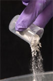
-This article was provided by Brookhaven National Laboratory.-Caption: SAMMS is a simple, inexpensive and easy-to-use technology that absorbs mercury in liquids and can be easily disposed of afterwards.
Pacific Northwest National Laboratory (PNNL) has developed an innovative technology that quickly and easily reduces or removes mercury content without creating hazardous waste or by-products, and that can be disposed of as a nonhazardous waste.
SAMMS™ (Self-Assembled Monolayers on Mesoporous Supports) is simple, inexpensive and easy to use; it is highly adaptable for use in reducing and removing metal contaminants from aqueous and non-aqueous materials; and it has numerous applications, including water treatment, waste stabilization, and metal processing and finishing. It is also significantly faster, more effective, and far less expensive than other mercury removal methods used in the past.
The SAMMS technology was first licensed to Steward Environmental Solutions, LLC, a manufacturer of advanced powders and nanomaterials. Steward signed its first licensing agreement in 2005, intending to initially market SAMMS for treating gaseous emissions such as those that come from coal-fired power plants, municipal incinerators, and other similar plants where testing has begun.
In March 2006, Steward signed a second license agreement for the manufacture and sale of SAMMS for multiple fields of use. PNNL continues to refine and test new applications that will broaden the range of contaminants effectively treated by SAMMS. The company hopes to work with PNNL on the production of these applications; Steward plans to produce SAMMS on an industrial scale.
Additional technology transfer activities for SAMMS have engaged Perry Equipment Company (to remove mercury from "produced water" resulting from off-shore drilling) and Chevron (formerly Unocal, to remove mercury from crude oil).
The technology continues to garner international recognition, including features in numerous high-profile scientific, technical and trade publications, and nods from the scientific community including an R&D 100 award and recognition as a finalist in the environmental category in Discover magazine’s annual awards for technological innovation.
Associated Patent: 6326326
-This article was provided by Pacific Northwest National Laboratory.-Caption: A. Paul Alivisatos, LBNL
Quantum Dot Corporation, Hayward, CA, has announced the introduction of five new products based on nanotechnology licensed from the laboratory of Paul Alivisatos at LBNL. The new products use nanometer sized "quantum dots" as the light emitting component in fluorescent probes for use in biological imaging. Due to the unique light emitting properties of the inorganic quantum dots, their performance is superior to that of existing materials, which rely primarily on organic dye molecules.
Fluorescent labeling is a widely used tool in the biological sciences. Antibodies to which fluorescent dyes have been attached bind to targeted proteins, revealing their location when viewed under a high resolution microscope with laser illumination. The fluorescent dyes now in use however, "photobleach" under repeated laser scans; their light emission weakens and eventually stops. Earlier work in the laboratory of Alivisatos had shown that inorganic nanoparticle "fluorophors," such as cadmium selenide, are much more stable under prolonged laser illumination. They are also up to an order-of-magnitude brighter than conventional organic dyes. Furthermore, the size of the nanoparticles determines the wavelength (i.e. the color) of the emitted light and that size, in turn, is easily controlled in their solution-based synthesis. (MSD Highlight 99-7). Thus, a number of different sized dots, each targeted to a different cell structure, can be illuminated simultaneously to provide a multicolored view of the locations and interactions of these structures. Realizing the potential to make a robust and flexible fluorescent labeling scheme based on these materials, Quantum Dot licensed the LBNL technology.
For more information, visit http://www.lbl.gov/msd/PIs/Alivisatos/03/03_12_qdot.html.
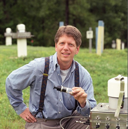
-This article was provided by Lawrence Berkeley National Laboratory.-Caption: Dr. Brian Looney, Senior Advisory Engineer, SRNL
Oxygen has long held promise as a means of remediating a variety of contaminants from groundwater, but effectively applying it underground has been problematic. A new method developed by Savannah River National Laboratory allows the permeation of a subsurface area with the right quantity of oxygen for effective groundwater remediation.
The solution, as developed by Brian Looney and Miles Denham, lies in the creation of metal peroxides in situ, which slowly react to release oxygen in a soluble form, providing a constant, relatively high concentration of oxygen dissolved in the groundwater. Metal peroxides do not exist naturally in soil, and placing them in the ground is often not practical, so Looney and Denham invented a way to convert metals and other materials already in place throughout the soil to peroxides or similar forms.
Their method involves injecting energetic oxidant solutions containing certain free radicals, which, when they interact with alkali earth metals like calcium and magnesium, form oxygen-rich peroxides that will release oxygen over time. These metals are commonly found distributed throughout the soil, making this a cost-effective way of applying peroxides.
Oxygen accomplishes remediation in various ways, depending on the contaminant involved. For organic contaminants, for example, oxygen can enhance the growth of natural microbes in the soil, which then use the contaminants as food sources, converting them to harmless by-products. Oxygen causes some metals to precipitate out of the groundwater, immobilizing them so that they will not be transported to downstream water sources. Other metals, such as chromium and uranium, become more soluble when exposed to oxygen; this allows them to be removed by filtration or other treatments.
This method is expected to be applicable to sites affected by a variety of different contaminants.
-This article was provided by Savannah River National Laboratory.-