|
|
[Last Modified: ] |
|
|
|
| [Trichomonas vaginalis] |
|
|
||||||
|
|
|
|
|
|
|
|
Microscopy
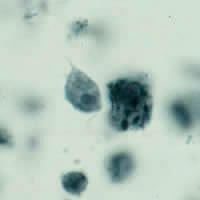
Trophozoites of Trichomonas vaginalis are pyriform and 7-30 µm long. They have five flagella: four anteriorly directed flagella and one posteriorly along the outer membrane of the undulating membrane. The large nucleus is usually located at the wider, anterior end and contains many chromatin granules and a small karyosome. The cytoplasm also contains many granules, but these are often not seen in Giemsa-stained specimens.
 |
 |
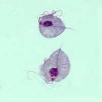
| A | B |
A: Two trophozoites
of T. vaginalis obtained from in vitro culture, stained with Giemsa.
B: Trophozoite of T. vaginalis stained with iron hematoxylin.
 |
| C |
C: Trophozoites of T. muris stained with iodine.
Click here to view a video clip #1 of Trichomonas
vaginalis (Adobe Flash)
Wet mount examination of vaginal
secretions from a patient with Trichomonas vaginitis. The organisms are
motile, pear-shaped, 10 µm by 7 µm, with visible flagella.
Click here to view a video
clip #2 of Trichomonas vaginalis (Adobe Flash)
Wet mount examination of a
culture of Trichomonas vaginalis.
|
|||