 |
Date Posted: October 3, 2008
|
|
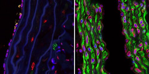
| Blood Vessels in Progeria Mice |
| Caption: |
Photomicrographs showing the blood vessel of an untreated mouse with progeria (left) compared to the blood vessel of a similar mouse treated with the farnsyltransferase inhibitor drug tipifarnib. The drug blocked loss of smooth muscle cells (stained green) in blood vessels, preventing the cardiovascular disease characteristic of progeria. |
| Credit: |
Michelle Olive, NHGRI |
| Additional Info: |
Confocal microscopy photographs of the descending aortas of two 15-month-old progeria mice, one untreated (left picture) and the other treated with the farnsyltransferase inhibitor drug tipifarnib (right picture). The microphotographs show prevention of the vascular smooth muscle cell loss that is otherwise rampant by this age. Staining was smooth muscle alpha-actin (green), lamins A/C (red) and DAPI (blue). (Original magnification, x 40) |
Date Created:
|
October 6, 2008
|
| Categories |
| Category (1): |
Science Photos: Other |
|
 |
|
|
