


About LANL
About Our Capabilities, Facilities, and Staff
"Los Alamos National Laboratory plays an indispensable role in building America as a science and technology powerhouse, and our staff are an incredible resource to the nation and the world." Michael Anastasio, Dir.
Solving Complex R&D Problems with Special Blend of Staff, Capabilities and Facilities
Now in its seventh decade, LANL remains among a very few laboratories that can bring great breadth of fundamental and discovery science, technology, and engineering rapidly together to create tangible solutions for national security needs.
Our staff, working with partners throughout science and industry, must be able to deliver today's solutions while maintaining the depth of capabilities to deliver the next generation of discoveries.
Los Alamos has demonstrated a cycle of innovation where we have developed world-leading capabilities and facilities in response to urgent, unique missions. We also spin out new discoveries that lead to emerging missions.
Being able to integrate and apply our capabilities rapidly to new challenges will be a key advantage in an increasingly competitive landscape.
Our Science, Technology and Engineering Priorities
Science that Matters
- Information science and technology enabling integrative and predictive science
- Experimental science focused on materials for the future
- Fundamental forensic science for nuclear, biological, and chemical threats
How We Work
- Collaborate, partner and team to make decisive contributions to our sponsors
- Outstanding operational excellence for safety, security, and efficient pursuit of ST&E for our missions
Transform Our Scientific Campus
- Campus for 2020 (consistent with complex transformation)
- Modern science facilities: LANSCE refurbishment, CMR replacement, Science Complex
- Signature facilities for experimental science (MaRIE) and computational science (Roadrunner)
More About This Science
Ultrasound-computed Tomography Uses a Prototype Scanning Device
Better Breast Cancer Detection
Safer, more comfortable and accurate tests find undetectable, highly fatal cancers

Quick read
We have developed a safer, more comfortable, inexpensive, and accurate way to detect early-stage cancer by using sound waves that replace dangerous x-ray technology used for mammograms.
Women may soon have access to safer, more comfortable, inexpensive, and accurate breast scans that find early-stage cancers. Breast cancer affects one in eight women in the U.S., and it is the second most common and fatal cancer.
Currently, the only routine breast-screening technology is mammography, an awkward and unpleasant procedure that uses x-rays to scan through tissue and capture on film a two-dimensional (2D) image of the breast.
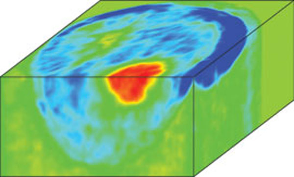
Los Alamos scientists Lianjie Huang and Kenneth M. Hanson and collaborators have developed a better way, producing a three-dimensional (3D) image, using sound waves instead of dangerous x-rays.
Utrasonic tests can detect early-stage breast cancers. Ultrasonic evaluation of breast lesions is desirable because it is quick, inexpensive, and does not expose the patient to potentially harmful ionizing radiation. Improved image quality and resolution enables earlier detection and more accurate diagnoses of tumors, thus reducing the number of biopsies performed, increasing treatment options, and lowering mortality and remission percentages.
X-ray mammography cannot accurately detect small tumors. Additionally, mammography subjects the patient to ionizing radiation, which carries inherent risks.
How it Works
The technique, called ultrasound-computed tomography (ultrasound CT), uses a prototype scanning device built at Karmanos Cancer Institute (KCI). A woman's breast is immersed in water and surrounded by a ring-shaped array of hundreds of ultrasound elements. Each element emits ultrasound waves and then receives waves that are scattered from the soft tissue. The array is moved incrementally down the entire breast, gathering data at each step.
A suite of newly developed computer algorithms converts the stepwise ultrasound data into a series of high-resolution, 2D images and then turns the series into a single 3D image. The technology actually obtains three kinds of images, corresponding to the speed, attenuation, and reflectivity of the waves.
Ultrasonic imaging takes advantage of differences in the interactions of acoustic waves with tissues of varying material properties. The interactions of the waves and the tissue cells are governed by wave-propagation physics and involve the following three phenomena: the speed of acoustic waves, scattering, and attenuation.
The Laboratory collaborated on this project with researchers from KCI, London's Imperial College, and Stanford University.
Ultrasound CT has the potential to detect cancer in its earliest stages. And since it is both safer and more comfortable, it should prove an attractive alternative for future breast cancer screening.
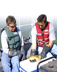
Robinson and Fresquez monitor air, soil, animals, plants
Data looks very favorable as the Lab continues working to mitigate its impact on the environment
Fresquez and his team are sampling lakes and rivers to determine the source and migration of potential contaminants.
Currents, the Laboratory's monthly employee magazine, highlighting people in the workplace.