|

Press Release 08-218
Next Generation Microscopy: No Stain, Big Gain

Researchers can monitor drug distribution and perform medical diagnostics rapidly using a new 3D imaging technique
December 18, 2008
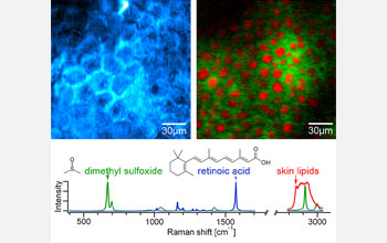
Microscopes have revolutionized the practice of science, especially in the fields of biology and medicine. Just a few hundred years ago, gaining the ability to study what was previously unobservable opened up an entirely new world. Today, imaging techniques remain indispensable to clinicians and researchers who regularly diagnose medical conditions and work to develop new treatments. Test results can often take hours or even days because cells or tissues must be subjected to lengthy fixation and labeling processes, sometimes called staining, in order to visualize and distinguish cellular components. In addition to long processing times, staining procedures often include harsh treatments or conditions that alter the tissues themselves, making interpretation of results difficult. A newly developed label-free imaging technique called stimulated Raman scattering (SRS) will likely revolutionize biomedical imaging in research and diagnostic laboratories. A team lead by Sunney Xie at Harvard University reported this new technique in the December 19 issue of Science. "It is a big step forward in terms of biology," said Xie. "SRS is a powerful imaging modality with widespread applications on many fronts of biology and medicine. This work compliments an earlier technique we developed with funding from the National Science Foundation, adding a new imaging modality to the vibrational microscopy field." The key to this new chemical imaging technique is the use of two lasers with different frequencies. Researchers visualize samples by tuning the laser frequencies to match the vibrational frequency of a specific chemical bond. Each type of molecule within a sample, including nutrients or drugs, is detectable at a unique frequency. By combining sample data collected at numerous frequencies, researchers can produce a high-resolution 3D image of the sample. SRS microscopy represents a big gain in biomedical imaging because it avoids labor-intensive sample preparation and autofluorescence, or "background noise", associated with traditional fluorescence microscopy. Xie is enthusiastic about the ways in which SRS imaging will facilitate progress in many fields. "Applications of SRS imaging range from mapping distribution of small metabolite and drug molecules in cells and tissues to medical diagnosis of cancer. Neuroimaging is another exciting area of application."
-NSF-

Media Contacts
Lisa Van Pay, NSF (703) 292-8796 lvanpay@nsf.gov
Lily Whiteman, NSF (703) 292-8070 lwhitema@nsf.gov
Principal Investigators
X. Sunney Xie, Harvard University (617) 496-9925 xie@chemistry.harvard.edu
The National Science Foundation (NSF) is an independent federal agency that
supports fundamental research and education across all fields of science and
engineering, with an annual budget of $6.06 billion. NSF funds reach all 50
states through grants to over 1,900 universities and institutions. Each year,
NSF receives about 45,000 competitive requests for funding, and makes over
11,500 new funding awards. NSF also awards over $400 million in
professional and service contracts yearly.
 Get News Updates by Email Get News Updates by Email
Useful NSF Web Sites:
NSF Home Page: http://www.nsf.gov
NSF News: http://www.nsf.gov/news/
For the News Media: http://www.nsf.gov/news/newsroom.jsp
Science and Engineering Statistics: http://www.nsf.gov/statistics/
Awards Searches: http://www.nsf.gov/awardsearch/
|