National Plan for Eye and Vision Research
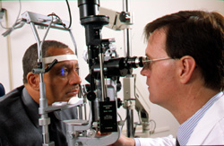
Glaucoma is a group of disorders that shares a distinct type of optic nerve damage that leads to loss of visual function. The disease is manifested as a progressive optic neuropathy that, if left untreated, leads to blindness. It is estimated that as many as 2.2 million Americans have glaucoma, and a similar number may have the disease without knowing it. Of these, as many as 120,000 are blind as a result. Furthermore, glaucoma is the number one cause of blindness in African Americans. Its most prevalent form, primary open-angle glaucoma (POAG), can be insidious. Unfortunately, since quality of life is not significantly affected until the later stages of the disease process, a significant proportion of individuals remain either undiagnosed or undertreated. Glaucoma usually begins in midlife and progresses slowly but relentlessly. If detected early, disease progression can frequently be arrested or slowed with drug and/or surgical treatment. A greater awareness of the deterioration of the optic nerve in the absence of elevation in the intraocular pressure or even functional field loss has reaffirmed the importance of recognizing the disease in its earliest stages. Continued laboratory and clinical research has provided greater understanding of the normal functions of the ocular tissues involved in glaucoma and other optic neuropathies. Past studies have led to a better understanding of glaucoma etiology and pathophysiology. The results have been the introduction of new glaucoma drugs with still more on the horizon, identification of a number of molecules and genes that play a role—either causative or secondary—in the disease mechanism, development of new diagnostic tools, clarification of the role of intraocular pressure (IOP), and introduction of new experimental disease models. However, there is still a significant need to improve methods of detecting structural and functional changes and develop new therapeutic options. More recently, a conceptual change has taken place within the glaucoma research community. Researchers now recognize that to understand glaucoma they need to understand the total neurodegenerative process, including the insults that initiate the neurodegenerative process, the mechanisms by which retinal ganglion cells die, and how these processes relate to end-stage optic nerve damage. In this regard, the workshop "Pathophysiology of Ganglion Cell Death and Optic Nerve Degeneration" was sponsored by the NEI to explore the state of knowledge of neurodegeneration as it applies to glaucoma. The full report Investigate the Pathophysiology of Ganglion Cell Death and Optic Nerve Degeneration can be accessed at http://www.nei.nih.gov/strategicplanning. On the basis of the recommendation from both this workshop and the NAEC that glaucoma be viewed as an optic neuropathy and be studied within that context, the Program will be expanded to include research on all optic neuropathies. The current overall emphasis for research in this Program is on identifying the biological mechanisms responsible for the various forms of glaucoma and other optic neuropathies so that improved treatment can be developed. The goals and objectives outlined in this Strategic Plan reflect these changes.
Over the past 5 years, results from three major clinical trials confirmed the value of reducing IOP in patients with ocular hypertension or glaucoma to prevent the onset of glaucoma in the former case and the progression of disease in the latter. The Ocular Hypertension Treatment Study (OHTS) noted that lowering IOP at least 20 percent produced a 50 percent protective benefit over baseline among those individuals who had elevated IOP without optic disc or visual field deterioration. The Early Manifest Glaucoma Trial determined that patients with newly diagnosed glaucoma progressed less often than untreated patients when IOP was reduced at least 20 percent compared with baseline. The Collaborative Initial Glaucoma Treatment Study demonstrated that patients with glaucoma who undergo either medical or surgical therapy were equally likely to avoid progression of disease after 5 years of followup. Analyses of key baseline, clinically important factors among ocular hypertensive patients enrolled in the OHTS uncovered or affirmed a number of risk factors for the development of glaucomatous damage, including IOP, large cup-to-disc ratio, age, and central corneal thickness.
Significant advances in identifying glaucoma-causing or associated genes have been made with the mapping of more than a dozen glaucoma loci and the cloning of more than a half dozen glaucoma genes. New studies involving genome-wide screening are beginning to identify alleles that may play a combinatorial role in complex POAG. Identification of trabecular meshwork glucocorticoid response/myocilin, optineurin, cytochrome P450 1B1 (CYP1B1), and other genes that play a less prominent role in disease causation promises a better understanding of normal eye development and of the molecular pathophysiology of glaucoma in general. The application of genomic technologies has provided an ever-enlarging database of genes/proteins expressed in various anterior segment tissues. Having a rich supply of candidate genes will speed the search for new genes involved in glaucoma pathogenesis. The introduction of a number of rodent models, both genetic mouse and induced ocular hypertension rat models, has expanded the ability to investigate mechanisms at the molecular and systems levels. One exciting new development in the field of congenital glaucoma is the recent report that tyrosinase modifies the glaucoma phenotype in the CYP1B1 knockout mouse. Significantly, this work supports findings that modifier genes play a role in the etiology of congenital glaucoma in children. Other models have allowed the evaluation of pharmacological approaches that target neurodegenerative processes. Over the past 5 years, candidates for molecular mediators of the pathophysiology of glaucoma have been identified. This list includes a number of second messengers, stress response proteins, immunologic proteins, and transcription factors. Myocilin was one of the first proteins to be associated with glaucoma. Much progress has been made on the characterization of myocilin, including an improved understanding of the differences in biochemical and cell biological characteristics between disease-causing and benign forms of the protein. New applications of technologies, such as the flash and multifocal electroretinogram and multifocal visual evoked potential, allow objective assessment of inner retina functioning in nonhuman animal models as well as in humans. Outcomes of both functional and histologic studies point to a pathological process that does not discriminate between subsets of ganglion cells and involves changes in the glial cells and retinal ganglion cell axons within the optic nerve head, as well as in the dendrites and somas of the retinal ganglion cells in the retina. Development of new and existing instrumentation for quantification of the retina, retinal nerve fiber layer, and optic nerve head surface has allowed substantial improvement in the clinical detection of structural damage. Algorithms for sensitive and specific screening (detecting glaucomatous damage) and change detection (monitoring glaucomatous progression) are approaching clinical usefulness for several of these instruments.
|