|
 |
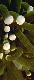
Major Reef-building Coral Diseases
 |
|
|
Sea fan with red band disease. (Photo: A. Bruckner)
|
|
|
Coral diseases and syndromes generally occur in response to biotic stresses such as bacteria, fungi and viruses, and/or abiotic stresses such as increased sea water temperatures, ultraviolet radiation, sedimentation and pollutants. One type of stress may exacerbate the other (Santavy and Peters, 1997).
The frequency of coral diseases appears to have increased significantly over the last 10 years, causing widespread mortality among reef-building corals. Many scientists believe the increase is related to deteriorating water quality associated with anthropogenic pollutants and increased sea surface temperatures. This may, in turn, allow for the proliferation and colonization of disease-causing microbes. However, exact causes for most coral diseases remain elusive. The onset of most diseases likely is a response to multiple factors (Peters, 1997).
This section discusses, in alphabetical order, the most prevalent coral diseases and syndromes currently known and under study: black-band disease, coral bleaching, dark-spots disease, red-band disease, white-band disease, white-plague disease, white pox and yellow-blotch disease. Additional information on these diseases and others can be found on NOAA's Coral Disease Identification and Information Web site.
Black-band Disease
Coral Bleaching
Dark Spots Disease
Red-band Disease
White-band Disease
White Plague
White Pox Disease
Yellow Blotch Disease
References
Black-band Disease
Black-band disease (BBD) is characterized by a blackish concentric or crescent-shaped band, 1 to 30 mm wide and up to 2 m long, that “consumes” live coral tissue as it passes over the colony surface, leaving behind bare skeleton. The disease is caused primarily by a cyanobacteria in combination with sulfide-oxidizing bacteria and sulfur- reducing bacteria, although other bacteria and opportunistic organisms such as nematodes, ciliate protozoans, flatworms and fungal filaments also are present in the mix (Richardson et al., 1997). The photosynthetic pigments of the dominant cyanobacteria gives the band its maroon to black color, but other bacteria are present in the mix. Often, the band has a white dusting of sulfur-oxidizing bacteria (NMFS, 2001).
 |
|
|
Black-band disease. (Photo: A. Bruckner)
|
|
|
The band, which is loosely anchored in the coral’s living tissue and is easily dislodged by water motion, advances across the surface of the coral from a few millimeters up to 2 cm per day. The unaffected coral tissue appears normal in color, morphology and behavior (McCarty and Peters, 2000). However, exposed coral skeleton is rapidly colonized by filamentous algae and other organisms (McCarty and Peters, 2000).
Black-band disease was first discovered on the reefs of Belize and Florida in 1972, and has since been identified in 26 countries including Fiji, Australia and the Philippines (Green and Bruckner, 2000). In the western Atlantic, BBD most commonly affects massive reef-building corals, but other types of stony corals and sea fans can be affected as well. Caribbean staghorn and elkhorn coral appear to be resistant to the disease (NMFS, 2001), though acroporid corals in the Red Sea and Indo-Pacific are affected by BBD. A total of 16 species have been observed with BBD in the western Atlantic, and 26 species in the Red Sea and Indo-Pacific (Green and Bruckner, 2000).
The number of corals infected with BBD on a reef fluctuates, but BBD is often present on most reefs at low levels, to depths of over 100 feet. The incidence of BBD increases in late summer and early fall, when water is clear and temperatures reach their peak (Green and Bruckner, 2000). The incidence and prevalence may also increase when corals are stressed by sedimentation, nutrients, toxic chemicals and warmer-than-normal temperatures (Richardson, 1998). Although colonies often suffer only partial mortality, loss of live coral tissue reduces the number of reproductive coral polyps and opens up surfaces that can be colonized by bioeroding organisms (Edmunds, 1991).
(top)
 |
|
|
Coral bleaching. (Photo: A. Bruckner)
|
|
|
Coral Bleaching
Healthy tissue of most stony corals ranges from yellow to brownish in color, a function of the photosynthetic pigments of their symbiotic zooxanthellae. When corals are inordinately stressed, they often expel their zooxanthellae, or the concentration of photosynthetic pigments declines. This response is known as bleaching (Glynn, 1996).
During a bleaching event, a coral’s coloration disappears or becomes pale, and the white of the coral skeleton shows through the translucent coral tissue. In some species, such as the massive starlet coral Siderastrea sidereal, the tissue can appear pinkish or bluish, due to pigments within the animal tissue. Localized bleaching has been observed since at least the beginning of the 20th century. However, beginning in the 1980s, regional and global bleaching affecting numerous species has occurred on reefs worldwide. Bleaching usually is not uniform over single coral colonies within coral communities or across reef zones, and some species are more susceptible to bleaching than others under the same conditions (Glynn, 1996). In some instances, only the upper surface or lower surface of the colony is affected. In others, bleached tissue appears as a circular patch or in the shape of a ring or wedge.
Localized bleaching has been attributed to exposure to high light levels, increased ultraviolet radiation, temperature or salinity extremes, high turbidity and sedimentation resulting in reduced light levels, and other abiotic factors (Glynn, 1996). In addition, bleaching in some species has occurred in response to a bacterial infection (Kushmaro et al., 1996). However, the seven major episodes of bleaching that have occurred since 1979 have been primarily attributed to increased sea water temperatures associated with global climate change and el Niño/la Niña events, with a possible synergistic effect of elevated ultraviolet and visible light (Hoegh-Guldberg, 1999).
Debilitating effects of bleaching include reduced skeletal growth and reproductive activity, and a lowered capacity to shed sediments and resist invasion of competing species and diseases (Glynn, 1996). Prolonged bleaching can cause partial to total colony death. If the bleaching is not too severe, and the stressful conditions decrease after a short time, affected colonies can regain their symbiotic algae within several weeks to months (Glynn, 1996).
 |
|
Dark-spots disease. (Photo: A. Bruckner)
|
|
|
Dark-Spots Disease
Though only recently described as dark-spots disease (DSD), discolored spots or markings in the tissue of several massive reef-building corals from the western Atlantic have been noted for many years, but not studied. Dark-spots disease was first reported from Colombia during the late 1990s, but the condition appears to be widespread in the Florida Keys and throughout the wider Caribbean (Gil-Agudelo and Garzón-Ferreira, 2001).
The affected areas appear as dark purple, gray or brown patches of discolored tissue, circular or irregular in shape, that are scattered on the surface of a colony, or at the colony’s margin. The discolored tissue increases in size and radiates outward as the area first affected dies. Darkened polyps often are depressed and appear smaller in size than normal polyps (Bruckner, 2001). DSD is most commonly observed on massive starlet coral (Siderastrea siderea) and blushing star coral (Stephanocoenia intersepta), but this condition also affects Montastraea annularis (species complex) (Bruckner, 2001).
 |
|
|
Close-up of red-band disease. (Photo: A. Bruckner)
|
|
|
Red-band Disease
Red-band disease (RBD) consists of a narrow band of filamentous cyanobacteria that advances slowly across the surface of a coral, killing living tissue as it progresses (Bruckner, 2001). Two types of RBD have been described. RBD-1 closely resembles BBD, but the band is reddish to maroon in color, and the cyanobacterial filaments are more loosely organized.
RBD-2 is visibly different from RBD-1, in that the cyanobacterial filaments spread like a net over the colony's surface (Richardson, 1992). Like BBD, RBD is dominated by filamentous cyanobacteria, forming a soft microbial mat that is easily dislodged from the surface of the coral tissue. However, different species of cyanobacteria have been found to be associated with RBD (Richardson, 1992; Santavy et al., 1996). RBD affects massive and plating stony corals, and also sea fans throughout the wider Caribbean. Like in BBD, exposed skeletal surfaces are rapidly colonized by algae and other competing organisms.
(top)
White-band Disease
White-band disease (WBD) was first identified in 1977 on reefs surrounding St. Croix. It is now known to occur throughout the Caribbean where it is believed to only affect staghorn and elkhorn corals (Green and Bruckner, 2000). This disease is characterized by tissue that peels or sloughs off the coral skeleton in a uniform band, generally beginning at the base of the colony and working its way up to branch tips (Peters, 1997). The band ranges from a few millimeters up to 10 cm wide, and tissue is lost at a rate of about 5 mm per day (Gladfelter, 1991).
 |
|
|
White-band disease. (Photo: A. Bruckner)
|
|
|
The effects of WBD can be devastating. In fact, WBD is thought to be a major factor in the decline of elkhorn and staghorn corals in the wider Caribbean (Aronson and Precht, 2001). Since the 1980s, Acropora cervicornis has been virtually eliminated from reef environments throughout the region. In the U.S. Virgin Islands, populations of Acropora palmata declined from 85 percent cover to 5 percent within 10 years, primarily as a result of WBD (Gladfelter, 1991). WBD currently is the only coral disease known to cause major changes in the composition and structure of reefs (Green and Bruckner, 2000).
Scientists have distinguished two forms of WBD. Type II, first identified on staghorn corals in the Bahamas in 1997, differs from type I in that tissue adjacent to exposed skeleton bleaches before it dies. Type II WBD sometimes is mistaken for bleaching (Ritchie and Smith, 1998).
The cause of WBD is unknown. Though unusual aggregates of rod-shaped bacteria were found in the tissue of corals affected by WBD type I, scientists have not determined the role of this microorganism. To further complicate matters, some corals that contain these bacteria appear healthy, and other colonies that are affected by sloughing tissue do not contain the bacteria (Richardson, 1998). More recently, scientists reported a species of bacteria associated with type II (Ritchie and Smith, 1998).
WBD has also been found throughout the Red Sea and Indo-Pacific, including the Philippines, the Great Barrier Reef and Indonesia. Unlike reports of WBD from the Caribbean, this condition has been identified on 34 species of massive, plating and branching corals in nine countries in the Indo-Pacific (Green and Bruckner, 2000).
 |
|
|
White plague disease. (Photo: A. Bruckner)
|
|
|
White Plague
White plague is similar in appearance to WBD, but it affects different species. The disease is characterized by an abrupt line or band of white, exposed coral skeleton that separates living tissue from algal-colonized skeleton, and often a narrow band of bleached tissue may be visible adjacent to exposed skeleton. Usually beginning at the base of a colony, it spreads quickly upward and outward.
White plague was first identified in the Florida Keys in 1977. A second form, type II, was identified on the same reefs in 1995, and a third form (type III) was reported in 2000 (Richardson and Aronson, in press). The three types of plague are similar in appearance, although a greater number of species are affected by type II. Additionally, the rate of tissue mortality is much greater in type II and type III than in type I (Richardson, 1998; Richardson and Aronson, in press).
Plague type I is reported to affect 10 species of corals, causing coral tissue mortality at a rate of about 3 mm/day. In Plague type II, up to 2 cm of tissue per day succumb to the disease, and small colonies can be decimated within one to two days. Thirty-two species are reported to be affected by this condition (Richardson, 1998). Plague type III affects the largest reef-building corals, including C. natans and M. annularis, and tissue loss is much greater than that observed in either plague type I or plague type II (Richardson and Aronson, in press).
 |
|
|
White pox disease. (Photo: A. Bruckner)
|
|
|
White Pox Disease
White pox affects elkhorn coral in the Florida Keys and possibly throughout the Caribbean. First found in 1996, the disease is characterized by white circular lesions on the surface of infected colonies. The rate of tissue damage appears to be rapid, allowing for algal colonization within days. The cause of white pox remains a mystery, but a possible bacterial pathogen has been identified (Porter, 2002).
(top)
 |
|
|
Yellow blotch /band disease. (Photo: A. Bruckner)
|
|
|
Yellow Blotch Disease
Affecting only star corals in the genus Montastraea and the brain coral Colpophyllia natans, yellow blotch disease (YBD) was first identified in 1994 in the lower Florida Keys. It is now known to occur throughout the Caribbean (Green and Bruckner, 2000). Yellow blotch disease begins as pale, circular blotches of translucent tissue or as a narrow band of pale tissue at the colony margin, with affected areas being surrounded by normal, fully pigmented tissue. As the disease progresses, the tissue first affected in the center of the patch dies, and exposed skeleton is colonized by algae (Bruckner, 2001). The area of affected tissue progressively radiates outward, slowly killing the coral.
The rate of tissue loss by corals afflicted with YBD averages 5 t 11 cm per year, which is less than that of other coral diseases. However, corals can be affected for many years, and the disease can affect multiple locations on a colony. Moreover, some of the largest and oldest coral colonies have been affected by YBD (Bruckner, 2001). Though the cause of YBD remains elusive, histological studies in affected corals have shown degenerative changes in tissues and cells. Scientists also have found unusual crystalline material in the coral's gastric cavity (Santavy and Peters, 1997).
References
Aronson, R.B. and W.F. Precht. 2001. Evolutionary palaeoecology of Caribbean coral reefs. In: Allmon, W.D. and D.J. Bottjer (eds), Evolutionary paleoecology: The ecological context of macroevolutionary change. NY: Columbia University Press. pp. 171-233.
Bruckner, A.W. 2001. Coral health and mortalit: Recognizing signs of coral diseases and predators. In: Humann and Deloach (eds.), Reef Coral Identification. Jacksonville, FL: Florida Caribbean Bahamas New World Publications, Inc. pp. 240-271.
Edmunds, P.J. 1991. Extent and effect of black band disease on a Caribbean reef. Coral Reefs 10:161-165.
Gil-Agudelo, D.L. and J. Garzón-Ferreira 2001. Spatial and seasonal variation of dark spots disease in coral communities of the Santa Marta area (Columbian Caribbean). Bull Mar. Sci. 69:619-630.
Gladfelter, W.B. 1991. Population Structure of Acropora palmata on the Windward Fore Reef, Buck Island National Monument, St. Croix, U.S. Virgin Islands. U.S. Virgin Islands: U.S. Department of the Interior, National Park Service. 172 pp.
Glynn, P.W. 1996. Coral reef bleaching: Facts, hypotheses and implications. Global Change Biology 2:495-509.
Green, E. and A. W. Bruckner. 2000. The significance of coral disease epizootiology for coral reef conservation. Biological Conservation 96:347-361.
Hoegh-Guldberg, O. 1999. Climate change, coral bleaching and the future of the world's coral reefs. Mar. Freshwater Res. 50:839-866.
Kushmaro, A., Y. Loya, M. Fine, and E. Rosenberg. 1996. Bacterial infection and coral bleaching. Nature 380:396.
McCarty, H. and E. Peters. April 1, 2000. The Coral Disease Page.
http://ourworld.compuserve.com/homepages/
mccarty_and_peters/Coraldis.htm.
National Marine Fisheries Service (NMFS), National Oceanic and Atmospheric Administration. 2001. Diseases of Reef-building Corals. http://www.nmfs.noaa.gov/prot_res/PR/
coraldiseases.html.
Peters, E.C. 1997. Diseases of coral reef organisms. In: Birkeland, C. (ed.), Life and Death of Coral Reefs. New York: Chapman & Hall. pp.114-139.
Porter, J. (ed.). 2002. Diseases in the marine environment. Hydrobiologia.
Richardson, L.L. and R.B. Aronson. In press. Infectious diseases of reef corals. Proc. 9th Intl. Coral Reef Symp., Indonesia.
Richardson, L.L. 1998. Coral diseases: What is really known? Trends in Ecology and Evolution 13:438-443.
Richardson, L.L., K.G. Kuta, S. Schnell, and R.G. Carlton. 1997. Ecology of the black band disease microbial consortium. Proc. 8th Intl. Coral Reef Symp. 1:597-600.
Richardson, L.L. 1992. Red band disease: A new cyanobacterial infestation of corals. Proc. 10th Ann. Amer. Acad. Underw. Sci. 153-160.
Ritchie, K.B. and G.W. Smith. 1998. Type II white-band disease. Rev. Trop. Biol. 46 Suppl. 5:199-203.
Santavy, D.L., T. Schmidt, S.S. Wilkinson, D H. Buckley and A.W. Bruckner. 1996. The phylogeny and alliance of cyanobacteria affiliated with two band diseases in corals from the western Atlantic. 96th ASM Meeting.
Santavy, D.L. and E.C. Peters. 1997. Microbial pests: Coral disease research in the western Atlantic. Proc. 8th Int. Coral Reef Symp. 1:607-612.
(top)
|
|
|
