|
New Targeted Fluorescent-Imaging Compound Allows Researchers to Detect Viable Cancer Cells in Mice Researchers have developed a new type of imaging compound that allows them to visualize viable breast cancer cells that have spread to the lungs in mice. The compound binds to a protein called HER2, which is found on the surface of some breast cancer cells, and it glows, or fluoresces, only when taken inside living cells. This method of targeting and activation allowed researchers to detect specific types of live cancer cells in a mouse model of breast cancer. The study, by researchers at the National Cancer Institute (NCI), part of the National Institutes of Health, and in Japan, appeared online Dec. 7, 2008, in Nature Medicine.
Previously developed fluorescent compounds that are activated inside the body's cells have the limitation that, once they are turned on, they continue to fluoresce even after they diffuse to new locations, making it difficult to distinguish viable tumor cells from normal tissue or dead or damaged tumor cells. The research team, led by Hisataka Kobayashi, M.D., Ph.D., at the Molecular Imaging Program of NCI's Center for Cancer Research (CCR), in collaboration with Yasuteru Urano, Ph.D., at the University of Tokyo, created an imaging compound that is turned on only when it is inside a living cell and stops fluorescing when it leaves the cell, as would happen when the cell dies or becomes damaged. The compound also can be engineered to target specific types of cancer cells.
"Imaging compounds designed by our concept may have applications in the clinic," said Kobayashi. "These compounds may allow clinicians to monitor a patient's response to cancer therapy by allowing them to visualize whether a drug hits its target and whether hitting the target leads to shrinkage of the tumor. With additional research and extensive testing in humans, these compounds may also be adapted for use in endoscopy procedures and for use as a surgical aid to improve removal of tumors."
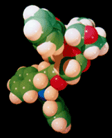
The team created the activatable, cancer-targeting compound by joining a drug called trastuzumab (Herceptin), which is an antibody that binds to HER2 and is used to treat HER2-positive breast cancer, to a modified version of a small fluorescent complex known as BODIPY. This complex fluoresces only under acidic conditions, such as those found inside cellular structures called lysosomes, which are sac-like compartments inside cells that contain enzymes that break down large molecules the cell does not need. When the activatable BODIPY-antibody compound encounters a HER2-positive breast cancer cell, the trastuzumab portion binds to HER2 proteins on the cell's surface, and then the cell takes the HER2-activatable complex inside. When this complex is processed inside the cell and enters the acidic environment of a lysosome, BODIPY becomes activated and fluoresces.
"Our design concept is very versatile and can be used to detect many types of cancer," said Kobayashi. "Unlike other activatable fluorescent compounds, our compound consists of a targeting agent and a fluorescing agent that act independently. We can target the fluorescing agent to different types of cancer cells by using any antibody or molecule that is internalized by the targeted cells after it binds to the cell's surface proteins."
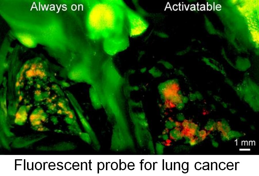
Using a mouse model, the Kobayashi team examined the potential of the activatable compound for detecting tumors within the body. They injected either the activatable compound, or a control that always fluoresces, into the tail of mice that had HER2-positive breast cancer tumors that had spread to their lungs. One day later, the investigators found fluorescence from the activatable compound only in lung tumors, whereas the "always on" control produced fluorescence in lung tumors, normal lung tissue, and the heart.
To confirm that the activatable compound was primarily processed by, or specific for, HER2-positive tumor cells, the researchers induced lung metastases in mice by intravenously injecting both HER2-positive tumor cells and tumor cells that carried the gene for red fluorescent protein (RFP) instead of the HER2 gene. After administering the fluorescence compounds, they found that the activatable compound produced fluorescence only in the HER2-positive tumors, whereas the "always on" control produced fluorescence in HER2-positive tumors and the surrounding tissues as well as the RFP-positive tumors.
Of the 472 HER2-positive tumors examined in mice with the activatable compound, only three showed fluorescence from both the activatable compound and the RFP (false positive), indicating that the activatable compound had a 99 percent tumor detection accuracy, or specificity, for HER2-positive tumors. The "always on" control had a specificity of less than 85 percent.
The team also confirmed that the activatable compound detects only living cells. Thirty minutes after they exposed tumor tissues to alcohol to kill the cells, they found that the fluorescence of the activatable compound significantly decreased in tumor tissue, whereas the fluorescence of the "always on" control showed minimal changes.
In another series of experiments, the researchers demonstrated the versatility of their design concept by linking a BODIPY complex to a molecule that targets the surface of mouse ovarian cancer cells. This compound allowed the researchers to detect clusters of live ovarian cancer cells that had spread to the peritoneum, or the tissue lining the walls of the abdomen, of mice.
###
Urano Y., Asanuma D., Hama Y, Koyama Y, Barrett T, Kamiya M, Nagano T, Watanabe T, Hasegawa A, Choyke P.L., and Kobayashi H. Selective molecular imaging of viable cancer cells with pH-activatable fluorescence probes. Nature Medicine. Online December 7, 2008.
For more information on Dr. Kobayashi's research in NCI's Molecular Imaging Program, please go to http://ccr.cancer.gov/labs/lab.asp?labid=175.
NCI leads the National Cancer Program and the NIH effort to dramatically reduce the burden of cancer and improve the lives of cancer patients and their families, through research into prevention and cancer biology, the development of new interventions, and the training and mentoring of new researchers. For more information about cancer, please visit the NCI Web site at http://www.cancer.gov or call NCI's Cancer Information Service at 1-800-4-CANCER (1-800-422-6237).
Back to Top |