All Images

Press Release 08-220
Viruses, Start Your Engines!

Researchers find what drives one of nature's powerful, nanoscale motors
Back to article | Note about images
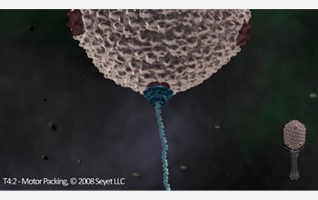 |
Image of DNA entering the gp17 motor complex on the T4 capsid. The image is a still from a video that can be found at: http://www.seyet.com/t4_academic.html.
Credit: T4:2 - Motor Packing, © 2008 Seyet LLC |
Download the high-resolution JPG version of the image. (68 KB)
|
Use your mouse to right-click (or Ctrl-click on a Mac) the link above and choose the option that will save the file or target to your computer.
|
 |
 View video View video
Michael Rossman, professor of biological sciences at Purdue University, describes his research on how viruses work. His interest is in the mechanics of viruses--the individual components of viruses and how they work together. How do molecular motors work and how is the mechanical energy of the fuel, ADP, transformed into motion.
Credit: Purdue University/The Catholic University of America/Seyet LLC
|
 |
View video
An animation showing bacteriophage T4 packaging DNA into its "head".
Credit: Purdue University/The Catholic University of America/Seyet LLC |
 |
View video
In a video from prior research in 2004, the bacteriophage T4 is preparing to infect its host cell. The structure of bacteriophage T4 is derived from three-dimensional cryo-electron microscopy reconstructions of the baseplate, tail sheath and head capsid, as well as from crystallographic analyses of various phage components. The baseplate and tail proteins are shown in distinct colors.
Credit: Purdue University and Seyet LLC |
|