Laboratory of Cardiac Energetics
NADH Fluorescence Image
|
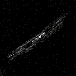
This is a NADH fluorescence image of a single living cardiac myocyte taken with a confocal microscope (optical slice thickness of 1uM, excitation 351nm, emmision around 450nm). The fluorescence comes from NADH located in mitochondria which are densely packed in myocytes. The molecule NADH plays an important role in aerobic mitochondrial respiration and the study of this molecule in living cells gives insight into how aerobic respiration is controlled in muscle. The cross hatch pattern that segregate the areas of NADH fluorescence are the t-tubules which do not fluoresce. The two large round non-fluorescent areas are cell nuclei. |