|

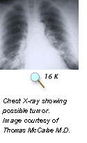
X-ray imaging is perhaps the
most familiar type of imaging. Images produced by X-rays are due to the
different absorption rates of different tissues. Calcium in bones absorbs
X-rays the most, so bones look white on a film recording of the X-ray image,
called a radiograph. Fat and other soft tissues absorb less, and look gray. Air
absorbs least, so lungs look black on a radiograph. The most familiar use of
X-rays is checking for broken bones, but X-rays are also used in cancer
diagnosis. For example, chest radiographs and mammograms are often used for
early cancer detection or to see if cancer has spread to the lungs or other
areas in the chest. Mammograms use X-rays to look for tumors or suspicious
areas in the breasts.
< Previous | Next Section > Main |