


|
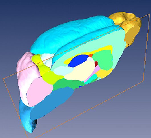
Sagittal cut through the 3D microMRI C57BL/6J mouse brain atlas The image shows in great detail a sagittal cut through the 3D microMRI C57BL/6J mouse brain atlas. Brain structures are labeled in different colors. The structures visible here are neocortex (cyan), olfactory bulb (golden), cerebellum (pink), brain stem (blue), caudate putamen (light green), hippocampus (deep blue), thalamus (milky white), external capsule (yellow), internal capsule (red), globus pallidus (deep pink), amygdala (light yellow), inferior coliculli (yellowish green), superior coliculli (white) and hypothalamus (deep cyan). Image courtesy of Dr. Helene Benveniste and Dr. Yu Ma, Brookhaven National Laboratory and Stony Brook University. Grant NIH R01 EB 00233-04 and P41 RR16105. Image courtesy of Dr. Helene Benveniste and Dr. Yu Ma, Brookhaven National Laboratory and Stony Brook University. Grant NIH R01 EB 00233-04 and P41 RR16105.
|

 |
 |
Department of Health and Human Services |
 |
National Institutes of Health |
 |
