  |
|
Structure & Function Research GroupHeparan Sulfate sulfotransferases catalyze the transfer of a sulfuryl group from PAPS, the universal sulfate donor, to the acceptor substrates heparan sulfate and heparin. These enzymes are found in the Golgi, and are responsible for dictating the various roles heparan sulfate plays in biological systems. Sulfation at specific hydroxyls and amines along the polysaccharide chain confers specificity for binding in various processes such as organogenesis, blood coagulation, growth factor/cytokine action, lipid metabolism, and viral infection. For example, sulfonation by 3-O-sulfotransferase isoform 1 on its substrate provides the required sulfate group on heparan sulfate to allow for antithrombin binding to thrombin and thus inhibition of blood coagulation. However, 3-O-sulfotransferase isoform 3 is involved in the biosynthesis of an entry receptor for Herpes Simplex Virus 1. To understand the substrate specificity of these enzymes the group has determined their structure in collaboration with Jian Liu at the University of North Carolina in the Department of Medicinal Chemistry and Natural Products. In collaboration with the Negishi lab in the Laboratory of Reproductive and Developmental Toxicology, the group has also solved crystal structures of two of the glycosyltransferases responsible for heparan sulfate biosynthesis, human glucuronyltransferase I (GlcAT-I) and mouse1,4-N-acetylhexosaminyltransferase (EXTL2) a member of the exostosin gene family. Both of these enzymes have been solved in the presence of the product donor substrate UDP, and the acceptor substrate as well as in the presence of the correct donor substrates. These structures provided the first structural insight into the mechanism by which heparan and condroitin glycosaminoglycans are synthesized. 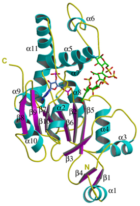 Figure 1: Ribbon diagram of the crystal structure of 3-O-sulfotransferase isoform 3 in complex with PAP (blue) and a tetrasaccharide substrate (green).
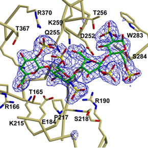 Figure 2: Electron density for the tetrasaccharide substrate (Moon et al. J. Biol Chem, 279:45185-93(2004)).
|
|