The Macromolecular Structure Group has used structural and biochemical approaches to understand how proteins specifically recognize their RNA targets. These studies have revealed the diversity of RNA recognition strategies that exist in nature.
Understanding Protein: RNA Recognition
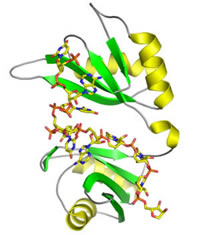
HuD recognition of AU-rich element RNA
Figure 1: Crystal structure of HuD RRMs 1 and 2 in complex with AU-rich element RNA.
Hu proteins regulate messenger RNA (mRNA) stability by binding to adenosine-uridine (AU)-rich elements (AREs) in the 3' untranslated regions of some mRNAs. These AREs have been shown to confer instability on the transcripts that contain them and are important players in regulating gene expression. Xiaoqiang Wang, a former postdoctoral fellow in the group, determined structures of an ARE-binding fragment of the HuD protein bound to a fragment of the c-fos ARE and to a fragment of the tumor necrosis α ARE. These structures identified residues important for RNA recognition and confirmed a consensus RNA recognition sequence for Hu proteins. Comparison to structures of other proteins containing RNA recognition motif (RRM) domains revealed a common binding mode for a number of RRM domains.
Post-transcriptional gene regulation by Puf proteins
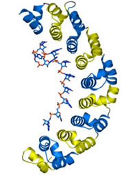
Figure 2: Crystal structure of the RNA-binding domain of human Pumilio1 protein in complex with target RNA.
Puf family proteins are found in organisms from humans to yeast and contain a conserved RNA-binding domain, the Pumilio-homology domain (PUM-HD). Puf proteins bind RNA sequence specifically and regulate translation and stability of target mRNAs. Xiaoqiang Wang, a former postdoctoral fellow in the group, determined the crystal structure of the PUM-HD from the human Pumilio1 protein alone and in complex with RNA. The protein comprises eight α-helical repeats and each of the eight repeats of the domain binds specifically to one RNA base using three side chains at conserved positions. As the pattern of side chains interacting with the bases was examined, a possible 'code' for RNA recognition was noted. In collaboration with Phillip Zamore’s lab at UMass Med School, this code was used to design a mutant protein where the three side chains in a single repeat were mutated to alter the RNA-binding specificity.
Engineering RNA sequence specificity of Pumilio1 protein
Using the code for RNA recognition deduced from crystal structures of human Pumilio1, Cheom-gil Cheong in the group has further probed the feasibility of creating Pumilo1 mutants with designed RNA binding specificity. An additional seven soluble mutant proteins with predictably altered sequence specificity were created, including one that binds tightly to AU-rich element RNA. These mutant proteins require only mutation of two base recognition residues in each repeat and bind their cognate RNA with high affinity. Thus Pumilio1 can be used as a scaffold to engineer RNA-binding proteins with designed sequence specificity. The group seeks to use this knowledge to design proteins as research tools that can be used to probe cellular function.
Mechanism of RNA silencing
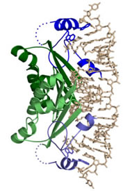
Figure 3: Crystal structure of CIRV p19 protein in complex with siRNA.
RNAi, the destruction of mRNA by double-stranded RNA containing corresponding sequences, has proven to be a powerful experimental tool and may have therapeutic potential. As a first step toward providing structural and biochemical insight into the mechanism of RNAi, in collaboration with Jozsef Burgyán’s lab at the Agricultural Biotechnology Center in Hungary, the laboratory has been studying p19, a plant viral protein that suppresses RNAi by binding to the double-stranded RNA intermediates, siRNA (small, interfering RNA). Jeff Vargason, a former postdoctoral fellow in the group, determined the crystal structure of the p19 protein from Carnation Italian Ringspot Virus in complex with a siRNA and examined the binding specificity of p19 for siRNAs, examining the features of siRNAs that allow recognition by this family of proteins. This structure suggested that the recognition of siRNAs is sequence independent and based on the size of the RNA duplex region. Binding experiments confirmed that p19 binds with highest affinity to RNA duplex regions of 18-20 nts and 30-300 fold more weakly to shorter or longer regions. p19 protein also inhibits microRNA function in plants, thus its binding to imperfectly matched duplex RNAs is being examined. The group is also studying proteins involved in processing small interfering RNA and microRNA.
Protein-facilitated RNA splicing
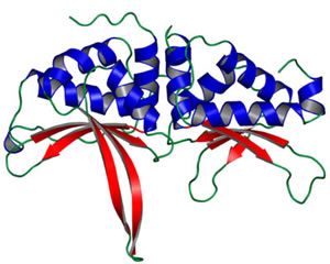
Figure 4: Crystal structure of the bI3 maturase from S. cerevisiae
LAGLIDADG homing endonucleases are widely dispersed in nature and bind across adjacent DNA major grooves via a saddle-shaped β-sheet recognition surface to catalyze DNA cleavage. Some LAGLIDADG proteins, called maturases, have been co-opted as splicing factors for select group I intron RNAs, raising the issue of how a DNA binding protein and an RNA evolve to function together. In collaboration with Kevin Weeks’ lab at the University of North Carolina, the crystal structure of the bI3 maturase was determined, which shows that the global architecture is unchanged from its DNA-binding homologs. The DNA endonuclease active site of the bI3 maturase, no longer necessary for activity as a splicing factor, has been concisely compromised with a single lysine group replacing a catalytic aspartyl-metal ion interaction. The bI3 maturase has been shown to bind to a peripheral group I intron domain at least 50 Å from the splicing active site. Protein-facilitated splicing by the bI3 group I intron thus represents a striking example of long distance structural communication in a ribonucleoprotein complex. Finally, site-directed cleavage and functional group interference experiments place the bI3 maturase nucleic acid recognition saddle at the RNA minor groove. Evolution from DNA to RNA function by the bI3 maturase has thus been mediated by a switch from major to minor groove interaction.