|
If you or someone you know has had a mammogram to check for breast cancer, an X-ray to detect a broken bone, or an ultrasound to examine an internal organ, then you’ve seen the benefits of medical imaging firsthand. Researchers and doctors can now see how the body works in whole new ways. Today’s powerful imaging tools help them understand diseases at more fundamental levels, which opens up new treatment strategies. It’s easy to see why medical imaging is such a rapidly growing field of research.
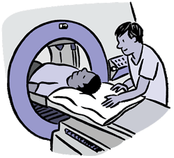
Illnesses and injuries are often treated more successfully and at lower cost when they’re detected early. That’s why imaging methods such as mammograms, CT scans and MRI are routinely used across the nation to spot cancer, heart diseases and other problems inside the body.
Mammograms, for example, can reveal abnormal areas within the breast before the woman or her doctor feels a lump. NIH’s National Cancer Institute predicts that, by the end of 2008, more than 182,000 women nationwide will be diagnosed with breast cancer and over 40,000 will die from it. Early detection of breast cancer is critical for survival. About 98% of women will survive for 5 years or more if their breast cancer is diagnosed before it spreads outside the breast. In fact, early detection by mammograms, along with improvements in treatment, has been driving down death rates from breast cancer since 1990.
Doctors recommend that women have regular mammograms once they turn 40, but some women are hesitant because of the discomfort involved. To address this problem, NIH-funded researchers have developed a new imaging technology, the breast CT, that’s more comfortable because the breast isn’t compressed during the procedure. The woman lies face down on a table and places one breast through an opening. The breast CT then uses X-rays taken from many different angles to develop a 3-dimensional image of the breast. The CT scanner gathers about 500 images as it rotates around the breast, so that researchers can look at the breast from any angle.
The breast CT is still being tested, but it may someday offer a pain-free way to spot cancer in breast tissue. It may also give doctors better insights into treatment options—for example, it could give them a more precise fix on abnormal areas. Researchers expect the device could be available within 3 to 5 years and become the new standard.
Other NIH research has brought changes in medical imaging that are already in practice. One example is a technology developed by NIH-funded investigators led by Dr. Richard Ehman at the Mayo Clinic. This technique, called MR elastography, sends gentle vibrations to the liver and uses MRI to evaluate the liver’s stiffness.
Doctors are now using MR elastography to assess scar tissue, or fibrosis, in the liver. The liver does many things in the body, including clearing poisons from your blood and helping control infections. Fibrosis of the liver can often be treated if it’s detected early. But if it’s not treated, it can progress to cirrhosis, an irreversible condition in which scar tissue replaces normal, healthy tissue and blocks the flow of blood through the organ, preventing it from working.
Typically, doctors take a biopsy to assess fibrosis of the liver. They use a needle to take a tiny sample of liver tissue and then examine it under the microscope for scarring and other signs of disease. MR elastography has now emerged as an alternative to this uncomfortable and invasive procedure.
According to Ehman, MR elastography has already made a difference in patient care. Ehman described one patient who has taken a potent drug for the past 15 years to control a severe skin disease. The drug can cause liver damage and fibrosis, so anyone who takes it needs to have a liver biopsy every 2 years to check for problems. At the patient’s last visit to the Mayo Clinic, however, the doctors used MR elastography to assess the liver. They found no evidence of fibrosis. “Patients are now able to avoid the pain, risk and higher expense of an invasive biopsy because of this technology,” Ehman explained.
Imaging by itself is not a treatment, but it can help doctors and patients make better decisions about treatments. NIH research and imaging discoveries are already improving the way health care providers detect and treat disease. Future advances will help doctors find disease earlier, guide treatments and assess whether treatments are working. |