 |
Insights From a Broken Brain
Ruth Levy Guyer, Ph.D.
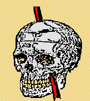
Explosive powders propelled a three and a half foot long iron rod into Phineas Gage's face. It pierced his left cheek, traveled behind his eye, and flew out the top of his skull. He didn't die. In fact, almost immediately after the accident, Gage could talk and walk.

Accounts of this bizarre accident along the railroad--Gage was a construction foreman--made the news bigtime in the spring of 1994 (1). That would not have been surprising considering the freakishness of the accident and its peculiar after-effects on Gage's personality, except for the fact that the accident occurred almost 150 years ago.
Gage had been tamping down dynamite on the roadbed. He and his crew were flattening the ground, preparing it for new train tracks. But he was working too fast: he pounded on the dynamite before his partner could cover it with a protective layer of sand. !!!POW!!! The rod became a rocket.
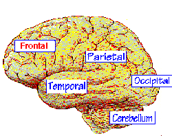
Gage's accident was newsworthy so long after the fact because brain researchers had just gained fresh insights into the nature of his injuries. They had carried out a detailed study of the pierced skull which, along with the rod, had been preserved by Gage's foresightful doctor and family, and they were able to tie together the structural damage to Gage's brain--specifically his frontal lobes--with his altered behavior (2,3).

In 1868, Gage's doctor, John Harlow, published a paper about his patient's remarkable recovery from the accident. This is the drawing that he included in his report. (See reference 4)
Before the accident, Gage had been a responsible, hard-working, fit and popular man. A report by his doctor that was published in 1868 (twenty years after the accident) described Gage as "a perfectly healthy, strong and active young man, twenty-five years of age, nervobilious temperament, five feet six inches in height, average weight one hundred and fifty pounds, possessing an iron will as well as an iron frame . . . having had scarcely a day's illness from his childhood to the date of this injury (4)."
After the accident, Gage became a nasty, vulgar, irresponsible vagrant. His former employer, who regarded him as "the most efficient and capable foreman in their employ previous to his injury," refused to rehire him because he was so different. "The equilibrium or balance, so to speak, between his intellectual faculties and animal propensities, seems to have been destroyed. He is . . . irreverent, indulging at times in the grossest profanity (which was not previously his custom), manifesting but little deference for his fellows, impatient of restraint or advice when it conflicts with his desires . . . obstinate, yet capricious and vacillating, devising many plans of future operation, which are no sooner arranged than they are abandoned in turn for others. . . "

But all had not been lost in Gage's brain. In fact, many of his intellectual skills were just fine, apparently untouched: "His mental operations being perfect in kind but not in degree or quantity (4)."
The researchers--Hanna and Antonio Damasio and their coworkers--combined photographs and x-rays of Gage's skull with computer graphics and produced on the computer screen a likeness of a brain that would have fit snuggly inside Gage's skull. Then they plotted the probable path of the flying rod through his head. From their analyses, it became clear that the rod had damaged not one but both of Gage's frontal lobes. That provided an explanation for the transformations to Gage's personality and behavior, because, although Gage's experience had been one-of-a-kind, its effects were not unique: other people with frontal lobe damage, including a number of the Damasios' patients, exhibit personality changes that resemble Gage's.

Twelve patients with frontal lobe damage, either brought on by traumatic injury or disease, are able, for example, as Gage was, to remember facts and perform complicated calculations. But, when it comes to keeping commitments, being trustworthy, holding a job, or succeeding in a marriage, they fail miserably. They can't plan for the future, and they can't see how their behavior will affect their own lives or the lives of others (1).
In their large registry of "broken brains," the Damasios have compiled data on more than 1500 specific brain lesions and the learning, memory, and personality changes that accompany them (3). The registry includes information about the brains of people who can pull up old memories but can't form new ones, those who can recall nouns but not verbs and others who can recall verbs but not nouns, and those who are unable to remember the names of other people or recognize faces, including their own. The brain of each of these patients, like an enticing and unguarded pie, seems to be missing a (functional) slice or two.

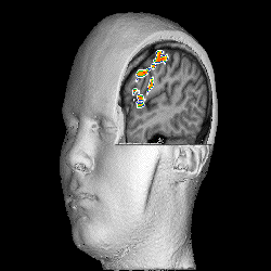
The subject was told to move his finger; his brain orchestrates the process. The picture here shows the regions of the brain that are participating in the process. Image courtesy of Dr. Peter Jezzard of the National Heart, Lung, and Blood Institute, NIH.
Two powerful brain "imaging" technologies--positron emission tomography (PET) and magnetic resonance imaging (MRI)--have provided pictures of intact and damaged brains for the brain registry. The raw PET and MRI data are converted into high-resolution maps of brains and brain lesions. These, in turn, provide fascinating and increasingly detailed clues to how the different parts of the brain contribute to the success of what seem like simple processes when they work properly--the recall of words, perception of colors, the ability to speak, name and face recognition, and so on.

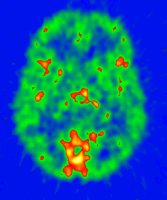
Remember a face. That's the task -- one that involves short-term memory -- that this person is doing. Specific regions become active as the brain works to remember the face, and these are the regions that are lit up in this positron emission tomography (PET) image. Courtesy of Dr. Susan Courtney of the National Institute of Mental Health of the NIH.
Studies of broken brains open windows onto healthy brains, because, by showing what can go wrong, such studies suggest what is going right in the brains of most people most of the time.
How a Positron Emission Tomography (PET) Scan Works
A person getting a PET scan lies down on a table. The part of the body -- say a section of the brain -- that is going to be studied is surrounded by detectors. The PET imager will be able to show, in three dimensions, where in the brain specific mental activities are taking place or where a tumor might be growing. The PET scanner picks these up, because, when a portion of the brain is active or when a tumor is growing, the flow of blood to that region is increased.
The detectors pick up radiation emitted from tagged (radioactive) compounds that are injected into the person's vein right before the scanner is turned on. The compounds travel in the bloodstream and arrive in the brain within a minute or so.
The tags used in PET scanning share one feature: they all emit positively charged electrons or "positrons." When a positron is emitted, it collides with an electron and forms a positronium. Both are "annihilated" by the collision, and their masses are converted to electromagnetic energy. Two energy packets in the form of gamma rays -- called 511-keV annihilation photons -- are released in opposite directions. They escape from the body, but the detectors pick them up. The detector determines that in fact two gamma rays have hit simultaneously 180 degrees apart. This is the signature of an annihilation, and from this signature the site can be mapped where the collision took place.
Positron-emitting "tags" for PET scanning are prepared in a cyclotron or a linear accelerator right before the radioactive compound is needed. The most common radioactive tags used for PET scanning are 18F, 11C, 13N, and 15O. After the positrons are emitted, the compound is no longer radioactive.
A three-dimensional PET image is compiled from a series of two-dimensional images or "snapshots" of the annihilation events. These events, in turn, indicate where brain cells are most active. A tumor will appear as a brighter-than-normal region. A thought or specific mental task will light up some region of the brain.
PET scans of brain regions involved in specific tasks are taken before the person performs the task (a measure of the steady state of blood flow in the brain) and during it. The difference in the two indicates what part or parts of the brain are involved in that activity. PET scanning is also useful for studying how the brain uses drugs, neurotransmitters, and other internal and injected substances.
How Magnetic Resonance Imaging (MRI) Works
A person who gets an MRI lies on a table that is then placed inside a device that produces a powerful magnetic field.
When the magnetic field is turned on, certain atoms in the body act like tiny magnets with north and south poles -- they line up in parallel columns that indicate the orientation of the imposed magnetic field. Then a series of radio wave pulses are delivered. These perturb the atomic "magnets," cause them to move, absorb energy, and, when the pulses are turned off, emit radio signals. The signals are characteristic of the atom that is emitting them. Through changes in the external field and the radio pulses, different atoms in the tissue can be activated.
About two-thirds of all atoms in the human body are hydrogen atoms. Hydrogen (1H) and 31P are the most common elements sought with MRI, but a lot of studies have also been done with 23Na, 31K, 19F, and 13C. All of these atoms share the property that they have either unpaired protons or unpaired neutrons or both in their nuclei. (Hydrogen has just one proton in its nucleus.) These unbalanced atoms wobble in the magnetic field and give off detectable radio signals.
MRI detects increases in oxygen in areas of heightened nerve cell activity. Its effectiveness reflects the way in which these cells use oxygen. It has the advantage that it does not depend on the injection of radioactive materials. With MRI, it is possible to detect tumors, chemical reactions, blood clots, and so on.
References:
- Science, May 20, 1994:1102. Many newspapers and magazines carried accounts of the Science report, including the New York Times Science Times (May 24, C1), the Washington Post (May 23, A2), and Science News (May 21, 326).
- Antonio Damasio, Descartes' Error, Gosset/Putnam, N.Y. 1994.
- Articles about the brain registry: Scientific American (September 1992, 89), Science, (May 18, 1990, 812), and the New York Times Magazine (October 18, 1992, 44).
- Harlow, John M. Recovery from the Passage of an Iron Bar Through the Head, 1868, Publication of the Massachusetts Medical Society, 2:327.
This article was originally posted on the NIH electronic bulletin board EDNET on 3/15/94. |