Contents
- Resource Brief
Modeling Brain Growth to Detect Disorders
Resource Brief
Modeling Brain Growth to Detect Disorders
With each visit to the pediatrician’s office, parents watch the doctor plot height, weight, and head circumference on growth charts. Although head circumference offers a general assessment of brain growth, it’s too crude a measure to offer insight into the structural and functional development of the brain.
That is why researchers at the Massachusetts General Hospital campus in Charlestown, Mass., home to the NCRR-funded Center for Functional Neuroimaging Technologies, are developing methods to model the development of the outermost layer of the human brain, the cerebral cortex. They may one day help doctors diagnose autism and other neurological disorders at an early stage by detecting clues about when and how the developing brain shows signs of abnormalities.
The developing human brain faces a basic “packaging” problem, according to Bruce Fischl, director of the neuroimaging center’s computational core, who led the research project published in the April 2007 issue of IEEE Transactions on Medical Imaging. The human cerebral cortex—the most recently evolved brain structure and one that plays critical roles in such complex functions as language, consciousness, memory, and perceptual awareness—is only a few millimeters thick but has the circumference of a medium pizza. “The only way to get the cortex to fit inside the skull is for it to fold,” Fischl says.
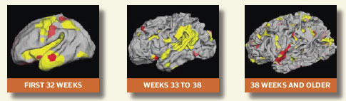
To create a mathematical model of the folding of the cerebral cortex during normal development, Fischl and Massachusetts Institute of Technology graduate student Peng Yu applied a mathematical analysis, called spherical wavelets, to cortical surfaces reconstructed from magnetic resonance (MR) scans of the brain. The cortical folding model, which was created using a group of eight newborns (several of them premature) and three children, predicts that the largest and deepest folds develop during the first 32 weeks of gestation, with folding accelerating during weeks 33 to 38 to complete smaller-scale folds. The smallest folds are predicted to develop at a faster pace, starting at 38 weeks of gestation. “These findings fit nicely with current observations of cortical folding development,” says Ellen Grant, chief of pediatric radiology at Massachusetts General Hospital in Boston. Grant and colleague Rudolph Pienaar are leading a large project to characterize, in detail, the cortical folding process.
To fit inside the skull, the developing human cortex has to undergo several levels of folding. Researchers have developed a mathematical model to predict the rate at which different folds normally develop. Once further refined, the model could help researchers detect neurological abnormalities even before clinical symptoms arise.
The model provides a proof of principle that the development of cortical folding can be precisely calculated. Once further refined, it could provide, along with additional measures, a more precise indication of brain growth and development than is offered by measurements of head growth. The hope is that such information will, in turn, allow earlier and more accurate determination of any deviations from normal brain development, possibly even before any clinical symptoms arise.
—Lisa Chiu
To Gain Access: The primary mission of the Center for Functional Neuroimaging Technologies is to expand understanding of the human brain in health and disease through the development and dissemination of innovative multimodal MR-based neuroimaging techniques and technologies. To learn more or to submit a proposal, visit the Center’s Web site. The Center is part of the NCRR-funded Biomedical Informatics Research Network (BIRN), a geographically diverse virtual community of shared resources to advance the diagnosis and treatment of disease.