|
National Institutes of Health |
|
|
|
Quick Links
|
Resource for Quantitative Functional MRION THIS PAGE: SEE ALSO: Resource for Quantitative Functional MRI
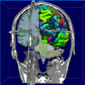
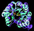
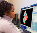
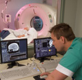
Research EmphasisThe resource is an interdepartmental and interdisciplinary effort consisting of four technical research and developments (TRD) projects and many collaborations dedicated to design novel magnetic resonance imaging (MRI) and magnetic resonance spectroscopy (MRS) data acquisition and data processing technology to facilitate the biomedical research of a large community of clinicians and neuroscientists at several institutions in Maryland and throughout the United States, with a special focus on pediatric and neurodevelopmental applications. The overall aims are to: 1) develop functional and physiologic MRI and MRS methods that can ultimately be used in a fast and quantitative comprehensive multimodality exam to assess pediatric disease; 2) develop image processing strategies to allow quantitative study of brain development applying the methods developed in AIM 1; 3) develop functional MRI processing strategies that are less dependent on subject compliance; 4) extend the presently available technologies for the brain as well as the to-be-developed technologies for application in the spine; 5) develop, apply, and validate these technologies in close collaboration with expert neuroscientists and clinicians; and 6) disseminate this new technology, train investigators in this new technology, and provide service to allow proper use of this technology. A big part of this effort is to provide these MRI technologies at 3 Tesla, and, in the near future, at 7 Tesla, where benefits of an increase in temporal, spatial, and spectral resolution can be exploited. TRD1: functional MRI (fMRI) data acquisition and analysis: fMRI analysis approaches that can reveal brain activity not predicted in advance by simple models (data-driven analysis), allowing extension of fMRI to the study of rich naturalistic behaviors and paradigms that require less subject compliance. TRD2: Multimodality MRI and MRS for functional assessment of brain and spinal cord pathologies. MRI and MRS approaches for quantitative determination of tissue physiologic and metabolic parameters. This includes new methods for blood volume MRI, magnetization transfer imaging, pH imaging, and spectroscopic imaging using parallel imaging approaches. TRD3: diffusion tensor MRI: High-resolution three-dimensional (3-D) diffusion tensor imaging (DTI) and fiber tracking/axonal mapping combined with multiple modalities of TRD2 for a tract-based physiologic analysis of brain and spinal cord. Together with TRD4, development of voxel-based analysis and tensor-based brain morphometry approaches to allow the study brain development. TRD4: algorithms for functional and anatomical brain analysis: Advanced computational technology, including Bayesian segmentation, volume-surface matching approaches, and dynamic tracking of vector fields. Methods for combining all image modalities into a general whole-brain reference frame. Current ResearchNew fMRI data analysis: Independent component analysis allows inferences from group, as opposed to single-subject data. Physiology and spectroscopy: Developed fast spectroscopic imaging using SENSE and new methods for blood volume weighted imaging without contrast agent and for pH-weighted imaging. Diffusion tensor imaging: High-resolution 3-D fiber tracking in humans using SENSE at 1.5 and 3 Tesla. First mapping of cortico-cortical association fibers and brainstem fiber and first in vivo white matter atlas. Elucidating specific fiber properties using MRI relaxation times and magnetization transfer. Computational anatomy: Validation of Bayesian segmentation algorithms for reconstruction of gyral areas. Derivation of probability laws for DTI data, resulting in dynamic programming algorithm for DTI tract tracing. Optimization of fMRI activation volumes. BIRNThe center is an active partner in the Biomedical Informatics Research Network (BIRN) of NCRR. Resource CapabilitiesThe resource combines facilities of the F.M. Kirby Research Center for Functional Brain Imaging at the Kennedy Krieger Institute and the Center for Imaging Science (CIS) at Johns Hopkins University. The Kirby Center has whole-body short-bore 1.5 T and 3 T Philips scanners and is being extended with a 7 T platform. CIS has an IBM supercomputer. A mock scanner for training children and difficult populations for functional exams is also available. SoftwareMultiple software packages are available online for data analysis of MRS and DTI data and our functional anatomy, and independent component analysis software has been implemented in other packages. Available ResourcesMultiple databases are available online, e.g., for pediatric brain DTI and group data for certain diseases (TRD4) and the software described above. Publications
|
| National Institutes of Health (NIH) Bethesda, Maryland 20892 |
Department of Health and Human Services |