

Home > News and Events > NIBIB Newsletter
In This Issue
LinksNational Institute of Biomedical Imaging and Bioengineering (www.nibib.nih.gov) Contact UsScience HighlightsNew Biosensor Measures the Interactions Between Biological Molecules Researchers supported by NIBIB have developed a new method to study the behavior of molecules, particularly how they interact with each other. In a study appearing in the September 21 issue of Science, chemists at Vanderbilt University, led by Darryl Bornhop, Ph.D., report that a new and deceptively simple technique—shining a red laser like those used in barcode scanners into a microscopic, liquid-filled chamber where two kinds of molecules are mixed—can measure the interactions between free-floating biological molecules, including proteins, sugars, antibodies, DNA, and RNA. In fact, the researchers have demonstrated that the new technique, called back-scattering interferometry or BSI, is sensitive enough to detect the process of protein folding. The method represents an entirely new application of interferometry, a powerful technique that combines light from multiple sources to make precise measurements. Interferometry is used in everything from astronomy to holography to geodetic surveys to inertial navigation. The equipment required for BSI is surprisingly modest: a helium-neon laser like those used in grocery store scanners, a mirror, a charge-coupled device or detector like those used in digital cameras, and a special glass microfluidic chip. The chip contains a channel about one fiftieth the size of a human hair. A “Y” at one end allows the simultaneous injection of two solutions, each containing a different kind of molecule. The “Y” is followed by a serpentine section that mixes the two solutions, followed by a straight section where the interactions are measured. An unfocused laser beam is directed through this section and reflected back and forth inside the channel approximately 100 times. Each time the light beam strikes the channel, some of the light is transmitted to the mirror, where it is directed to the detector. There it forms a line of alternating light and dark spots called an interference pattern. It turns out that the interference pattern is very sensitive to what the molecules are doing. If the molecules begin sticking together, for example, the pattern begins to shift. The stronger the binding force between the molecules, the larger the shift. This allows the system to measure interaction forces that vary a million-fold, which includes the entire range of binding forces found in living systems. The underlying physics of this highly sensitive measurement technique are still being worked out. The researchers know that it responds to minute changes in the index of refraction, a measure of how fast light travels through the liquid in the chamber compared to its speed in a vacuum. They hypothesize that this results from the rearrangement of water molecules that cover the surface of the proteins: When two proteins react they squeeze water molecules out of the area where they bind together. This displacement changes the density of the liquid slightly, which in turn alters its index of refraction. Vanderbilt has applied for and received two patents on the process and has several other patents pending. The University has issued an exclusive license to develop the technology to Molecular Sensing, Inc. Darryl Bornhop, Ph.D., is one of the founders of the startup and serves as its Chief Scientist. The company plans on completing a prototype system this fall. Additional information including images can be found at www.vanderbilt.edu/exploration/stories/backscatter.html. Reference: Microsurgery Device Reduces Surgeon Tremor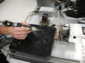 Microsurgical procedures require precision. Involuntary movement or jerk can traumatize surrounding tissue and cause myriad complications for the patient. A research team at Carnegie Mellon University has developed a handheld device that compensates for unwanted motion and stabilizes the tip of microsurgical instruments. The device could improve the safety of microsurgery and reduce practitioner fatigue. Clinical applications include retinal surgery, neurosurgery, cardiac surgery, and cell biology. To learn more about the work of Cameron Riviere, Ph.D., and other NIBIB grantees, please visit www.nibib.nih.gov/HealthEdu/eAdvances. NIBIB NewsNIBIB Awards Quantum GrantsNIBIB recently announced the award of more than $12 million in grants to support research and development of potentially high-impact, innovative technologies to advance health care. The new grants will fund four investigators in developing groundbreaking technologies: disposable microchips for the diagnosis of metastatic lung cancer, a bio-artificial kidney to eliminate dialysis procedures, insulin-producing cells to treat diabetes, and nanoparticles that selectively leave the blood and bind to cancer cells to assist in removal of brain tumors. “This innovative program from NIBIB promises to harness the power of technological discovery and team science to translate new knowledge into practical healthcare benefits for our nation,” said Elias A. Zerhouni, M.D., NIH Director. The overall goal of the NIBIB Quantum Grants Program is to make a profound (quantum level) advance in health care by funding research on targeted projects that will develop new technologies and modalities for the diagnosis, treatment, or prevention of disease. “We are excited to be awarding these Quantum Grants to four excellent researchers and their interdisciplinary teams,” said NIBIB Director Roderic I. Pettigrew, Ph.D., M.D. “We look forward to watching the extraordinary results that will be achieved as these studies progress. All four of these projects have the potential to significantly improve the current practice of medicine.” The four awardees are: Diabetes impacts the individuals afflicted and society as a whole due to the significant complications associated with using existing insulin treatment strategies. The aim of this project is to develop a new source of insulin-secreting cells as a replacement strategy for treating diabetes. Transplantation of pancreatic islets to restore insulin production is promising; however, donor pancreata are in short supply. The development of these tissue-engineered islets will provide a new source of insulin-producing cells and help realize the full potential of cell therapy for diabetes. Raoul Kopelman, Ph.D., University of Michigan at Ann Arbor, $2.6 million (3 years) Brain cancer, one of the most lethal forms of cancer, is diagnosed in more than 43,000 new patients each year. The goal of this project is to improve surgical resection and treatment options for brain cancer patients by developing nanoparticles that selectively leave the blood and bind to cancer cells. These nanoparticles will aid in the visualization of tumors to allow for maximal surgical resection of tumor mass and facilitate nonsurgical destruction of the residual cancer cells that are remote or extend from the tumor mass. This may achieve significant improvement in treatment of brain tumors. Shuvo Roy, Ph.D., Cleveland Clinic Lerner College of Medicine-CWRU, $3.2 million (3 years) End-stage renal disease is a significant global health problem. Donor kidneys for transplantation are in short supply, with dialysis and filtration as the only alternative treatment. A miniaturized, implantable, and self-regulating bio-artificial kidney that integrates the dialysis machinery into a miniaturized, implantable device will be developed. The successful development of this bio-artificial kidney would provide an alternative to the majority of the dialysis procedures performed annually in the U.S. Mehmet Toner, Ph.D., Massachusetts General Hospital, $3.4 million (3 years) The goal of this project is to develop a point-of-care microchip device that can determine the type, severity, and aggressiveness of a wide range of cancers by detecting tumor cells that are circulating in the blood stream. A disposable microchip technology capable of separating specific circulating tumor cells from whole human blood at concentrations as low as one in a billion will be developed. Detecting the presence of these tumor cells at such low concentrations enables earlier intervention in the treatment of metastatic lung cancer, which remains the leading cause of cancer death in the U.S. This point-of-care test can potentially transform patient care through early molecular diagnosis of lung cancer and identification of new biomarkers with which to track disease progression. For more information about the NIBIB Quantum Grants Program, visit www.nibib.nih.gov/Research/QuantumGrants. Division of Bioengineering and Physical Science Transferred to NIBIBNIBIB recently announced the integration of the Division of Bioengineering and Physical Science (DBEPS), formerly part of the NIH Office of Research Services, into the NIBIB Intramural Research Program.
This new intramural component has joined the existing NIBIB Intramural Research Program, which includes the PET Radiochemistry Research Laboratory responsible for conducting research and training in the development and application of novel radiochemical probes for biomedical imaging, and the joint Laboratory for the Assessment of Medical Imaging Systems at the FDA.  Multi-Agency Tissue Engineering Science (MATES) Strategic PlanThe MATES Interagency Working Group has released a strategic plan for Federal Government investments in tissue science and engineering, including the activities of both research and the regulatory agencies with interests related to tissue engineering or regenerative medicine. The plan, entitled “Advancing Tissue Science and Engineering, A Foundation for the Future,” was drafted by members of the MATES Interagency Working Group and reviewed and approved by the Office of Science and Technology Policy and the National Science and Technology Council. The strategic plan is available online at tissueengineering.gov/welcome-s.htm. NIBIB and India Partner to Develop Low-Cost Medical TechnologiesNIBIB and the Department of Biotechnology (DBT) of the Ministry of Science and Technology of the Republic of India have entered into a bilateral agreement to develop low-cost healthcare technologies aimed at the medically underserved. The agreement is based on a shared commitment to improve the health and well-being of the people of both countries by encouraging collaboration and cooperation on the development of diagnostic and therapeutic medical technologies that are inexpensive and operate at the initial point of physician contact, or point of care. "We are very pleased to officially establish this groundbreaking effort between NIH/NIBIB and the Department of Biotechnology," said NIBIB Director Roderic I. Pettigrew, Ph.D., M.D. "This agreement will create a working partnership designed to help address global health disparities by encouraging the development of improved methods and technologies to diagnose and treat illness and injury across geographic and economic borders." Areas of cooperation outlined in the agreement include low-cost innovations in x-ray technology; nanotechnology-based biosensors; point-of-care diagnostic technologies; telehealth and telecommunication technologies; and neonatal health technologies. The disease areas and conditions likely to be affected by the successful development of the technologies are infectious diseases, cardiovascular disease, liver disease, trauma and injury, and conditions associated with infant mortality. "Developing low-cost health technologies that are unique in design to be affordable and useable in disease prevention and management is a high priority in India," said Maharaj Bhan, M.D., DBT Director. "The partnership with NIH, and through them, with U.S. institutions, is critical for us to make progress. We are excited about this agreement with NIH to bring multiple disciplines and teams together to find innovative solutions." 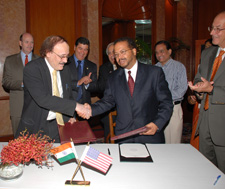 As part of the agreement, NIBIB and the DBT will encourage workshops and meetings to share experiences and scientific information; link appropriate Centers of Excellence and Institutes; engage in bilateral cooperation on the assessment and application of new diagnostic technologies; and generate collaboration among scientists and engineers in the conduct of research, research training, and technology development. The agencies will facilitate and share each other's efforts in research and development through regular interactions between scientists, and will work towards mutual, annual goals. The signing of the Agreement on Science and Technology took place during a recent visit to the Ministry of Science and Technology in New Delhi, India.
NIBIB Creates Point-of-Care Technologies Research NetworkNIBIB's new Point-of-Care Technologies Research Network funds four centers working to build expertise in the development of integrated systems that address unmet clinical needs in point-of-care (POC) testing through the creation of multidisciplinary partnerships. Specifically, each will serve as a resource to identify clinical needs in POC testing and communicate these needs to technology developers to better guide device design and appropriate clinical testing. Opportunities for collaboration on exploratory technology development projects will be announced as each center defines clinical needs in its respective health care area. Each center was awarded a grant via the U54 cooperative agreement mechanism. The specific goals and research interests of each center are described briefly below. The Center to Advance POC Diagnostics for Global Health, led by Dr. Bernhard Weigl—Program for Appropriate Technology in Health (PATH)—will accelerate the development and deployment of POC diagnostic tests essential to low resource centers by conducting laboratory and field-based evaluation on selected prototype POC diagnostic tests. Specifically, the center will create a device to measure CD4+ cell counts for AIDS treatment management as well as a multiplexed device to diagnose malaria, HIV, and syphilis in pregnant women. The Point-of-Care Center for Emerging Neurotechnologies, led by Dr. Fred Beyette—University of Cincinnati—will develop technologies to improve the diagnosis and response to neurodiseases and neurodisorders, particularly in emergency scenarios. Specifically, the center is developing and testing a spectrophotometric device for diagnosis of subarachnoid hemorrhage in ER patients. The Center for Rapid Multi-pathogen Detection for Point-of-Care Technologies and National Disaster Readiness, led by Dr. Gerald Kost—University of California, Davis—will improve the accessibility, portability, and field robustness of POC devices for critical care units, emergency rooms, community hospitals, rural areas, and disaster response sites. Specifically, the center will focus on simultaneous detection of multiple pathogens in human whole-blood samples, with reduction to practice as one prototype for hospital settings and a second more robust prototype for rapid field diagnosis in emergencies and disasters. The Center for Point-of-Care Technologies for Sexually Transmitted Diseases, led by Dr. Charlotte Gaydos—Johns Hopkins University—will create and test unique methods for diagnosis of sexually transmitted diseases, including the home delivery of OTC assays to end users via the Internet. Additionally, the center will develop novel approaches for measuring acceptability and accuracy of OTC-type assays in primary care settings with comparisons between trained and untrained users. The creation of the Point-of-Care Technologies Research Network came in response to recommendations from the April 2006 workshop, Improving Health Care Accessibility through Point-of-Care Technologies sponsored by NIBIB, the National Heart Lung and Blood Institute, and the National Science Foundation. This workshop assessed the clinical needs and opportunities for use of POC technologies in primary care, the home, emergency medical services, and in developing countries, and highlighted state-of-the-science sensors and microsystems, low-cost imaging, noninvasive monitoring, and telehealth/informatics. Funding Opportunities & UpdatesArmed Forces Institute of Regenerative MedicineThe U.S. Army Medical Research and Material Command, with the Office of Naval Research, and the NIH are establishing the Armed Forces Institute of Regenerative Medicine (AFIRM), dedicated to the repair and regeneration of battlefield injuries through the use of tissue engineering and regenerative medicine. Therapies developed by AFIRM will also serve trauma and burn patients in the public at large. The closing date for applications was October 19, 2007. The full funding announcement can be found at www.grants.gov/search/search.do?oppId=15133&mode=VIEW or at www.usamraa.army.mil/pages/index.cfm (click on PAA). NIH Roadmap for Medical ResearchThe NIH Roadmap is a series of far-reaching initiatives designed to build on the progress in medical research achieved through the doubling of the NIH budget. NIBIB plays a significant role in Roadmap activities in many areas. More information on current NIH Roadmap funding opportunities is available at nihroadmap.nih.gov/grants/index.asp. NIH Blueprint for Neuroscience ResearchThe NIH Blueprint for Neuroscience Research is a cooperative effort among the 16 NIH Institutes, Centers, and Offices that support neuroscience research. By pooling resources and expertise, the Blueprint supports the development of new tools, training opportunities, and other resources to assist neuroscientists in both basic and clinical research. More information on current NIH Blueprint funding opportunities is available at neuroscienceblueprint.nih.gov/blueprint_funding/index.htm. Names in the NewsNew Faces at NIBIBDr. Zeynep Erim has joined the Division of Interdisciplinary Training as a Program Officer. AwardsNIBIB Staff Roderic I. Pettigrew, Ph.D., M.D., NIBIB Director, has been elected to the Institute of Medicine (IOM). Members are elected through a highly selective process that recognizes people who have made major contributions to the advancement of the medical sciences, health care, and public health. Election is considered one of the highest honors in the fields of medicine and health. Current active members elect new members from among candidates nominated for their professional achievement and commitment to service.
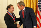
GranteesTwo NIBIB grantees were recently awarded the National Medal of Science by President George W. Bush at a White House reception:
Jacqueline A. Johnson, Ph.D., from Argonne National Laboratory recently received an R&D 100 Award for an Ultra-High-Resolution Mammography System (UHRMS). The UHRMS equips doctors with a low-cost, high-quality alternative to digital radiography.
Robert G. Griffin, Ph.D., from the Francis Bitter Magnet Laboratory, Massachusetts Institute of Technology was awarded the ENC’s Günther Laukien Prize for 2007 for “successful development of high-field dynamic nuclear polarization (DNP) for sensitivity enhancement in solid-state NMR with magic-angle spinning.”
John P. Donoghue, Ph.D., from Brown University has received the 2007 K.J. Zülch Prize, Germany’s top neuroscience award for “pioneering BrainGate, the mind-to-movement device that allows people with paralysis to control assistive devices using thoughts alone.” Conferences & MeetingsUpcoming Meetings and ExhibitionsSPIE Medical Imaging
AIMBE 17th Annual Meeting - The Global Impact of Medical and Biological Engineering PITTCON 2008 The NIH CornerEnhancing Peer Review at NIHThe two-tiered review process is the cornerstone of the system used by NIH to support biomedical and behavioral research. NIH Director Elias Zerhouni, M.D., has called upon leaders from across the scientific community and NIH to join a trans-NIH effort to examine the two-level NIH review system. The goals of this review are to optimize the system’s efficiency and effectiveness and ensure that the NIH will be able to continue to meet the needs of the research community and the public-at-large. Detailed information on the review is available at enhancing-peer-review.nih.gov. |


 |
 |
Department of Health and Human Services |
 |
National Institutes of Health |
 |
