News and Events
Congressional Justification for FY 2001
National Eye Institute
- Introduction
- Science Advances and Future Research Directions
- National Eye Health Program
- New Initiatives
- NEI Budget Policy
Authorizing Legislation: Section 301 and Title IV of the Public Health Service Act, as amended. Reauthorizing legislation will be submitted.
Budget Authority:
|
FY 1999 Actual |
FY 2000 Estimate |
FY 2001 Estimate |
Increase or Decrease |
||||
| FTE | BA | FTE | BA | FTE | BA | FTE | BA |
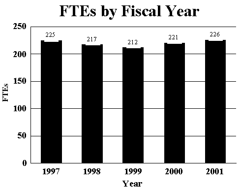
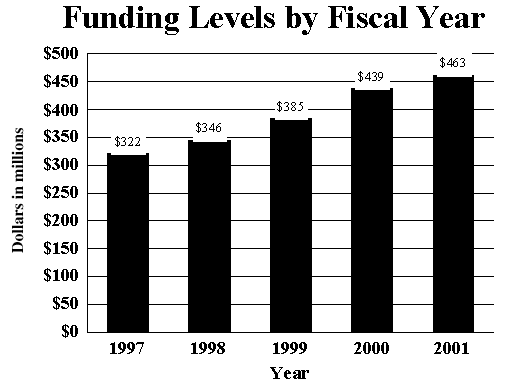
| 212 | $385,253,000 | 221 | $439,211,000 | 226 | $462,776,000 | +5 | +$23,565,000 |
This document provides justification for the FY 2001 Non-AIDS activities of the National Eye Institute. Justification of NIH-wide FY 2001 AIDS activities can be found in the NIH section entitled Office of AIDS Research (OAR).
Introduction
Out of its concern for the eyesight of the American people, Congress created the National Eye Institute (NEI) in 1968 with the mission to conduct and support research, training, health information dissemination, and other programs with respect to blinding eye diseases, visual disorders, mechanisms of visual function, preservation of sight, and the special health problems and requirements of the blind. Inherent in this mission is clinical research across the spectrum of diseases of the eye and disorders of vision, as well as the investigation of the normal tissue and normal visual processes that will help gain a more complete understanding of the abnormal processes that lead to these conditions. These investigations are conducted in hundreds of extramural laboratories and clinics throughout the United States and in the NEI's own intramural facilities in Bethesda, Maryland. The highlights that follow are examples of the research progress that is being made with the investment of federal funds in NEI-supported research and the direction that research will take over the next year.
Science Advances and Future Research Directions
Retinal Diseases
The retina is the complex, light-sensitive, neural tissue in the back of the eye that contains highly-specialized and metabolically active photoreceptor cells (rods and cones). These cells respond to light by emitting chemical and electrical signals. These signals are received by other retinal cells which process and transmit visual information via the optic nerve to the brain for decoding. The choroid is the underlying layer of blood vessels that nourish the retina.
The retina and choroid are susceptible to a variety of diseases that can lead to visual loss or complete blindness. These sight-threatening conditions include age-related macular degeneration, diabetic retinopathy, retinopathy of prematurity, retinitis pigmentosa, retinal detachment, uveitis (inflammation), and cancer (choroidal melanoma and retinoblastoma).
Complications of Age-Related Macular Degeneration Trial (CAPT): The CAPT is a large clinical trial involving 1000 patients and 24 clinical eye centers across the United States. The study will determine whether the use of low intensity laser treatment can prevent the advanced complications of age-related macular degeneration (AMD) and the associated vision loss. Over the next two years, 1000 patients with early signs of AMD, who also meet the other eligibility criteria, will be enrolled in the study. All patients will have one eye treated and one eye not treated. Both eyes will be watched closely for 5 years so that the effects of the CAPT laser treatment can be critically analyzed and evaluated. This study will increase our knowledge of AMD and may result in improved treatment that will prevent the devastating vision loss that can occur with AMD.
Retinal Transporter Proteins: A molecule, called the rim protein (RmP), of the retinal rod photoreceptor cell is a component of the outer segment discs. RmP was discovered in 1978 but no function could be assigned to it for many years. Recently, this protein has been associated with a specific eye disease--Stargardt's disease or fundus flavimaculatus. The gene for Stargardt's disease has been identified and shown to produce a "transporter molecule" that moves substances into and out of the cell. Recent work has demonstrated that RmP is identical with the transporter molecule responsible for Stargardt'sdisease. In Stargardt's disease, a pigment called lipofuscin accumulates in the pigmented epithelium, a layer of cells that underlie and nourish the retina. Lipofuscin is composed mainly of a molecule called A2E. In animal models in which the transporter gene has been deleted or "knocked out" scientists found that A2E levels were much higher than normal. So it appears that if this transporter protein is functioning improperly, A2E accumulates in the photoreceptor discs, leading to subsequent dysfunction. Further study should yield a better understanding of the pathogenesis of photoreceptor cell dysfunction that may ultimately help prevent or effectively treat retinal degenerative diseases.
Growth Factor Protects Nerves and Halts Blood Vessel Growth: Pigment epithelium-derived growth factor (PEDF) is a protein found in the healthy eye. PEDF is secreted by the retinal pigment epithelial cells that underlie and nourish the neural retina. Recently, a team of scientists demonstrated that PEDF can transiently delay the death of photoreceptors in mouse models of inherited retinal degenerations. This protein has also been shown to promote neurite-outgrowth and protect spinal cord motor neurons against natural and induced death using cell culture and animal model systems. Another group of scientists has shown that PEDF can prevent the growth of endothelial cells that form new blood vessels. Thus, PEDF behaves as a potent neurotrophic factor for the retina and nerves of the central nervous system, as well as a potent inhibitor of angiogenesis. Continued research to learn how PEDF works may provide information that will contribute to the development of effective treatments for several neural degenerative and angiogenic diseases, such as retinitis pigmentosa, macular degeneration, and diabetic retinopathy.
Adaptive Optics: Researchers supported by the NEI have shown that it is possible to image the three photoreceptor cone types in the intact living retina with a technology called adaptive optics. Adaptive optics technology was originally developed for astronomy: telescopes equipped with adaptive optics permitted the capture of clear images despite a turbulent atmosphere. Recently, this technology has been applied to the visual system, giving the clearest views yet of the living retina inside the eye. Adaptive optics allow one to have a much sharper image of the retina than previously possible, to the point of resolving single cells. One can apply a technique called retinal densitometry in combination with adaptive optics to identify a photopigment type unique for each cone. Since this technology is noninvasive, it may now be possible to examine the integrity of photoreceptors and other cell types in the living eye, to track the progression of a number of retinal diseases such as retinitis pigmentosa, or evaluate the efficacy of rescue of cell types in the retina. Research will continue to examine these possibilities.
Beneficial Effects of Oxygen in Retinal Detachment: Retinal detachment is a condition in which the sensory retina separates from its normal position lining the back of the eye near the capillary bed, which is the primary source of nutrients and oxygen. Scientists have recently found that animals with induced detached retinas demonstrated less retinal cell death and greater cell integrity when maintained in a high oxygen environment for several days. Since oxygen treatment can be easily administered in physicians" offices and hospitals, further research in this area may lead to a new approach to delaying the progression of cell damage in retinal detachment in humans.
Drug Inhibits Growth of New Blood Vessels in the Eye: A new therapeutic agent has been developed that may be important in treating blindness in humans caused by diabetic retinopathy or macular degeneration. Vessels that grow abnormally in the eyes can leak fluid or blood, causing rapid and severe vision loss. The new drug, called PKC 412, can be taken orally and appears to have several actions on growth factors and their receptors within the retina. While PKC 412 blocks new abnormal vessel growth, it has no apparent adverse effects on normal, fully mature vessels. Additional research is needed to determine whether PKC 412 is a viable therapeutic alternative in the treatment of diabetic retinopathy.
Corneal Diseases
The cornea is the transparent tissue at the front of the eye that serves two specialized functions. The cornea forms a protective physical barrier that shields the eye from the external environment. It also serves as the main refractive element of the eye, directing incoming light onto the lens. Refraction depends on the cornea acquiring transparency during development and maintaining this transparency throughout adult life. Corneal disease and injuries are the leading cause of visits to eyecare clinicians, and are some of the most painful ocular disorders. In addition, approximately 25 percent of the American population have a refractive error known as myopia or nearsightedness that requires correction to achieve sharp vision1.
Mechanism of Recurrent Ocular Infection by Herpes Simplex Virus (HSV-1): Following an initial eye infection by HSV-1, a lifelong latent infection of ocular nerves can result in periodic reactivation of the virus, recurrent disease, and potential blindness. Scientists have recently learned that one gene, LAT, is active during latency and enhances reactivation. Neuron survival is clearly important for latency of HSV-1, and researchers recently discovered that LAT inhibits programmed neuronal cell death in infected cells. Not only do these results provide important new information on neuronal survival, but they may also provide meaningful clues to researchers studying ways to inhibit the reactivation process.
Ocular Estrogen Receptor:Gender-based differences in the incidence of several important ocular conditions raise the possibility that estrogens may have direct effects on the human eye. Scientists using immunochemical techniques recently detected estrogen receptor protein in the retina and retinal pigment epithelium of young females, but not in eye tissues of men or postmenopausal women. This hormone receptor may offer a new therapeutic target for the treatment of dry eye syndromes.
Lens and Cataract
Cataract, an opacity of the lens of the eye, interferes with vision and is the leading cause of blindness in developing countries. In the U.S., cataract is also a major public health problem. Approximately 1.35 million cataract surgical procedures were performed on Medicare beneficiaries alone in 1991 costing approximately $3.4 billion.2 Cataract surgery accounts for approximately 12 percent of the entire Medicare Part B budget and is the most commonly performed surgical procedure.3 The enormous economic burden of cataract will worsen significantly in coming decades as the American population ages. The major goals of this program, therefore, are to determine the causes and mechanisms of cataract formation, to search for ways to slow or prevent the progression of cataract, and to develop and evaluate new diagnostic and therapeutic techniques in cataract management.
Lens Fiber Cell Replication and Differentiation: The lens of the eye is an ellipsoid-shaped structure that helps focus images onto the retina. The lens consists of only two cell types: epithelial cells, which form a single layer on the outer surface, and mature fiber cells that fill the internal cavity. Fiber cells are made from the lens epithelial cells in a process called differentiation. In order to maintain the number of epithelial cells, each epithelial cell replicates itself before it goes on to become a fiber cell. Scientists are attempting to identify the elements that direct the epithelial cell to stop replicating and start differentiating. These researchers have identified the gene product of Prox1, a gene which is expressed in a variety of embryonic tissues, that appears to be a central regulator of the transition from replication to differentiation. Studies in mice indicate that Prox1expression regulates the expression of elements known as cell cycle inhibitors that are required to stop the replication of epithelial cells and begin their differentiation into fiber cells. Absent Prox1 expression, cell cycle inhibitors are not expressed, causing replication without subsequent differentiation and fiber cell formation. This process can cause a complication that often occurs after cataract surgery known as "secondary cataracts," a situation in which residual cells begin replicating and differentiating in an attempt to form a new lens. If this process interferes with vision, laser treatment is required, thereby increasing treatment costs. It is essential to understand the regulation of replication and differentiation of these cells in order to devise new cost-effective preventive strategies.
Lens Protein Modifications in Cataract: Aging-related cataract is the leading cause of world blindness. At present there is no non-surgical means of prevention or treatment. A primary mechanism for loss of transparency of the lens involves aggregation of lens proteins that absorb or scatter light. Understanding the mechanisms underlying this aggregation process is critical for development of an effective medical therapy for cataract. Scientists have identified in certain human cataracts two atypical, modified forms of one of the major lens proteins, B-crystallin. The concentration of this protein is also increased significantly in these cataracts. It is one of a family of proteins thought to protect cells against stresses such as heat shock, osmotic stress, and oxidative stress by preventing the aggregation of proteins, an effect that would be particularly beneficial in the lens. The identification of specific modifications to lens proteins associated with cataract development will help understand the etiology of the disease and increases the possibility that this discovery could be an important step toward the development of an effective cataract treatment in the future.
Cataract Formation: Access to genetic tools has offered fresh approaches to understanding cataracts. Scientists recently discovered that a mutation in a human gene encoding a protein required for cell-to-cell communication causes congenital cataracts. When a mutant form of this gene is introduced into mice, the mice are born with cataracts. Because this protein'sstructure and function have been characterized, research can now be focused on developing a better understanding of the pathogenesis of cataracts with the goal of preventing their occurrence.
Glaucoma
Glaucoma is a group of eye disorders which share a distinct type of optic nerve damage that can lead to blindness. Elevated intraocular pressure is frequently, but not always, associated with glaucoma. Glaucoma is a major public health problem and the number one cause of blindness in African Americans. Approximately three million Americans have glaucoma4, and as many as 120,000 are blind from this disease5. Most of these cases can be attributed to primary open angle glaucoma, an age-related form of the disease. NEI activities in glaucoma research are directed toward understanding the mechanisms of the disease through basic research, identifying risk factors, and preventing blindness.
Neuroprotection in Glaucoma: Elevated intraocular pressure is frequently associated with glaucoma and explanations for how axons become damaged are usually based on the mechanical effects of elevated intraocular pressure. However, optic nerve damage can occur without abnormally high pressures and conversely, elevated pressure does not necessarily lead to optic nerve damage. Discovering the basis of optic nerve degeneration is essential for the development of the next generation of glaucoma drugs, neuroprotective agents. Scientists now have evidence that the molecule nitric oxide (NO), is directly involved in mediating the degeneration of axons in the optic nerve head. Using a rat model of glaucoma in which intraocular pressure is artificially elevated, scientists first found that rat optic nerve pathology mimics that of humans. Treating these rats with a compound that inhibits the production of nitric oxide protected the axons of these rats even though intraocular pressure remained higher than normal. By showing a connection between the production of a physiological agent, nitric oxide, and damage to retinal nerve cell axons, the results of this research are the first to suggest a pathway. Research is now being aimed at identifying and developing neuroprotective agents as a new class of glaucoma drugs.
Strabismus, Amblyopia, and Visual Processing
Developmental disorders such as strabismus (misalignment of the eyes) and amblyopia (commonly known as "lazy eye") affect 2-4 percent of the United States population6 ,7. The correction of strabismus is a frequently-performed surgical procedure. In addition to research relevant to strabismus and amblyopia, the NEI supports investigations of the age-related inability of the lens to focus on nearby objects, irregular eye movements, and refractive errors. Three million Americans now have chronic visual conditions that are not correctable by eye glasses or contact lenses8. Therefore, the NEI also supports research on improving the quality of life of persons with visual impairments by helping them maximize the use of remaining vision and by devising aids to assist those without useful vision.
Visually-Guided Behavior: The decision to focus the eyes on one of two targets begins with the sighting of both targets (sensory input) and ends with the coordinated movement of the eyes to focus on one target (motor outcome). The milliseconds between sensory input and motor output are more than a simple "knee jerk" reflex response as once thought. Primates trained to respond to visual cues demonstrate that anticipation of a reward, based on past experience, or the relative size of the reward associated with the target greatly influences target selection. The decision- making process is correlated with the increased firing of neurons in the sensory-motor processing area of the central nervous system. This research provides an important framework to model and understand the neurophysiological basis of visual-guided behaviors during the next year.
Specificity in Visual Signaling Pathways: Signaling molecules can be organized into different pathways within the same cell. Assembling these signaling molecules into architecturally- defined complexes is emerging as an essential cellular mechanism to ensure the specificity and selectivity of signaling. Recently, an NEI-funded scientist found that a Drosophila protein, InaD, brings together several components of the phototransduction cascade into a macromolecular complex. This complex was found to be the underlying element allowing effective and specific signaling in the process of vision. Continuation of this research will provide a more complete understanding of the visual process in health and disease.
Organization of Neurons in the Cerebral Cortex: One of the major issues in understanding the function of the cerebral cortex of the brain is how the activity of populations of neurons are coordinated to produce a coherent output. NEI scientists studying single neurons in the monkey brain have found that active populations of neurons shift from an averaging mode to a winner- take-all mode as the visual stimulus conditions controlling eye movements change. These experiments show that as the neuronal activity changed, the type of eye movement generated also changed indicating that the type of movement generated can be accounted for by the shifts in overlapping activity of cortical neurons. This type of research is critical to providing greater insight into the functioning of the cerebral cortex, as well as other areas of the brain.
New Help for Patients with Congenital Nystagmus: Congenital nystagmus is a condition that begins at birth or early infancy where the eyes oscillate continuously and uncontrollably. This condition can be familial, but more commonly it is associated with many diseases of the visual system that also begin in infancy. Treatment of this condition is usually aimed at associated problems, e.g., strabismus, anomalous head posturing, refractive error correction, and amblyopia treatment. It has been recently discovered in an animal model of this condition that surgical cutting of the extraocular muscle tendons (endotomy) decreased the nystagmus and improved the visual behavior. This procedure has entered a phase I study in humans that will be continued during the next year with the hope of decreasing their nystagmus and improving the visual function of patients with this condition.
Mathematical Modeling of Rapid Eye Movements: When we read or look around, our eyes make rapid movements (called saccades) that point at objects of interest so that we may see them more clearly. If these saccades are not accurate, or if the eyes continue to drift after them, vision is significantly impaired. Many areas of the brain must cooperate to make accurate, drift-free saccades. Building mathematical models of the functions performed by these different areas allows us to understand how the brain converts visual information into an eye movement model. Additional research will provide the understanding that is essential in dealing with abnormal saccades caused by disease, drugs, trauma, or aging.
Health Disparities
Ocular Hypertension Treatment Study (OHTS):In its early stages, glaucoma is usually treated with drugs in daily eye drops. In some patients, the beneficial effect of the eye drops lessens with time, and "advanced glaucoma" develops. Recent findings from the Advanced Glaucoma Intervention Study suggested that black and white patients with advanced glaucoma respond differently to two surgical treatments for the disease. Although both groups benefit from treatment, scientists found that blacks with advanced glaucoma benefit more from a regimen that begins with laser surgery, while whites benefit more from one that begins with an operation called a trabeculectomy. However, the role of topical medications in preventing or delaying sight-threatening damage to the eye from glaucoma remains unclear. In order to evaluate the safety and efficacy of topical ocular hypotensive medications in preventing or delaying damage to the optic nerve and loss of vision from primary open angle glaucoma (POAG), the NEI has funded the OHTS. The OHTS is designed to determine the potential benefit of treatment with ocular hypotensive medications in preventing or delaying damage to the eye from glaucoma. Additionally, the high percentage of African Americans participating will also ensure adequate evaluation of the effectiveness of topical medication in treating African Americans with glaucoma as the study progresses toward conclusion in the coming years.
Los Angeles Latino Eye Study: The NEI is supporting a major research project in Los Angeles County, California, the Los Angeles Latino Eye Study, to gain a greater understanding of the prevalence and incidence of eye disease among Latinos. Researchers are conducting in-depth interviews with study participants on their medical and ophthalmic histories, use of medications, tobacco and alcohol consumption, and utilization of health care services. Because so little is known about the visual health needs of this segment of the population, the data collected from this study will be instrumental in determining the prevalence of cataract, glaucoma, age-related macular degeneration, and diabetic retinopathy among Latinos in this community. The study will also determine the proportion of blindness and visual impairment that is caused by these diseases, and will explore the association of various risk factors, such as smoking or sun exposure with ocular disease. The study will also examine the effect of eye disease and disorders on quality of life and will assess the cost/benefit of eye care services and the utilization of those services in the Los Angeles Latino community. Continuation of this study should yield results that will assist in setting eye and vision health services priorities in the Los Angeles area and the rest of the country.
Visual Impairment Among Hispanics in Arizona Study: Another study supported by the NEI that is designed to improve our understanding of eye disease and visual impairment in the Hispanic population in this country is the Visual Impairment Among Hispanics in Arizona Study. This study is collecting data to determine the prevalence of diabetic retinopathy, cataract, and other causes of blindness and visual impairment in 4,500 Mexican Americans age 40 and older residing in Arizona. The results will provide new information of visual impairment and blindness in the Mexican-American community.
Uveitis
Uveitis Treatment: Uveitis is an inflammation inside the eye that affects primarily children and young adults. If left untreated, uveitis can cause blindness. NEI intramural scientists conducted a study to evaluate the safety and potential usefulness of a monoclonal antibody that is directed against a receptor on immune cells. These receptors bind to a product of the immune system, interleukin-2. Interleukin-2 plays an important role in activating immune cells, and therefore plays a central role in an inflammatory response in the body. This antibody was given to patients with uveitis thought to be due to autoimmunity and not to an infection. All patients who entered the study needed systemic immunosuppressive agents to control the inflammation in their eyes, and to maintain relatively good vision. Once patients received the monoclonal antibody (Zenapax), they were slowly weaned off their standard immunosuppressive medications. Nine of the ten patients who entered the study were able to stop their regular medication, ultimately receiving antibody therapy once a month. The results suggest that anti-interleukin-2 receptor therapy may be an effective therapeutic approach to the treatment of uveitis, as well as other autoimmune conditions, with potential improvement of vision and quality of life for patients. This research will now be extended to a larger group and might also have application in other disorders that are autoimmune and non-infectious in nature, such as multiple sclerosis and psoriasis.
Modulation of Uveitis Through Feeding of Retinal Proteins: Feeding of retinal proteins to animals protects them from developing autoimmune retinal disease after a subsequent immunization with the same proteins, a phenomenon known as oral tolerance. Clinical studies suggest that a similar phenomenon may operate in humans. Scientists studied the involvement in this process of soluble factors, known as cytokines, that are produced by lymphoid cells and regulate their function. Studies with mice that were genetically engineered to lack particular cytokines indicated that two anti-inflammatory cytokines, known as interleukin-4 and interleukin-10, are involved in this process. Research is now being focused on determining whether augmentation of these cytokines may be a useful approach to potentiating oral tolerance in a therapeutic setting.
National Eye Health Education Program
Low Vision Education Program: On October 19, 1999, the NEI announced the formal launch of its Low Vision Education Program. Low vision is broadly defined as a visual impairment, not corrected by standard glasses, contact lenses, medicine, or surgery, that interferes with the ability to perform everyday activities. Most people develop low vision because of eye diseases, such as cataracts; glaucoma; diabetic retinopathy; or age-related macular degeneration, the leading cause of severe visual impairment and blindness in Americans 60 years of age and older. Low vision primarily affects the growing population of people over age 65 and other higher risk populations, including Hispanics and African Americans who are likely to develop low vision at an earlier age. While lost vision usually cannot be restored, many people can learn to make the most of the vision that remains. The Low Vision Education Program will include a multimedia public service campaign and a traveling exhibit that will be displayed in shopping malls around the country. The program also will provide communities nationwide with materials and technical support to increase awareness of local low vision services and resources.
New Initiatives
Many of the following initiatives are part of broadly-based initiatives that are trans-NIH in research scope and content and will be conducted in cooperation with other institutes. They are initiatives that will be pursued within the NIH areas of emphasis established by the NIH director in concert with the institute and center directors. The importance of these initiatives for vision research is highlighted below under the area of emphasis to which they refer.
Biology of Brain Disorders
Neurodegeneration Research: Research on neurodegeneration and the rescue and regeneration of neural cells is an area of tremendous opportunity with application to many neurological diseases and conditions, and to cases of traumatic injury. This includes:
- Rescue of Photoreceptors in Retinal Degenerative Diseases--A number of research advances now support the development of strategies for preventing or slowing down photoreceptor degeneration in retinal degenerative diseases. There are numerous opportunities for basic research in this area, as well as opportunities for translating these research advances to the clinic. A number of approaches show promise, including the use of growth factors, transplantation, ribozymes, and other methods of gene therapy.
- Survival of Retinal Ganglion Cells--Retinal ganglion cells (RGCs) can be highly purified and cultured in serum-free conditions. Trophic factors have been discovered that are necessary for survival and protection from cell death. However, RGCs are unresponsive to these trophic factors unless they are active electrically. These effects are mediated through an elevation of intracellular cAMP. RGCs in culture and co-culture conditions provide a special research opportunity for investigating signaling mechanisms that normally promote survival and how these mechanisms are altered by injury.
- Protection of Axons in the Optic Nerve Head--Researchers have found elevated levels of nitric oxide synthase in the optic nerve heads from human eyes with glaucoma and animal models of glaucoma. By pharmacologically inhibiting the production of nitric oxide in these animals, scientists found that axons of the optic nerve were protected from neurodegeneration.
Genetic Medicine
Identification of Genes Expressed in the Visual System:The initial focus will be a gene discovery phase to identify and sequence genes that are expressed in the visual system. High- quality cDNA libraries will be prepared from ocular tissue that are representative qualitatively and quantitatively of the genes expressed in the visual system, optimized to detect rare or unique sequences. The ultimate goal will be to generate a complete set of full-length cDNA clones and their sequences from the visual system, which provide more comprehensive information on the protein coding region. It is anticipated that this catalogue of genes expressed in the visual system will be publicly available in an easily accessible and retrievable format, including a collection of arrayed cDNAs representing mRNAs expressed in human and mouse ocular tissue.
Bioengineering, Computer, and Advanced Instrumentation
Development of Novel Instruments for Micro-imaging: Signals generated by existing and next- generation probes need to be detected and analyzed. In the last several years, a range of technologies has been developed that permits better spatial resolution of signals, better capabilities for understanding three dimensional structure, and better ways to follow molecular and cellular processes in time. Research activities in this area could include:
- Development of high speed multi-photon microscopy to track the movement, and quantitate the amount of probes reporting intracellular signal transduction processes and other molecular cascades.
- Research on instruments for spectral analyses of Fluorescent Resonance Energy Transfer data generated by intracellular processes.
- Design and development of software to analyze and compare datasets from imaging instruments.
- Studies of the relationship of invasively and non-invasively observed brain activity- related optical signals.
New Approaches to Pathogenesis
Research on Methods to Control Angiogenesis:Diseases that affect the retinal blood vessels are among the major causes of visual disability and blindness in this country. These include diabetic retinopathy, retinopathy of prematurity, neovascular glaucoma, and age-related macular degeneration in which the proliferation of abnormal blood vessels (neovascularization) can result in the rapid and irreversible loss of vision. Investigations by NEI-funded researchers into the cellular mechanisms involved in the development of new or abnormal blood vessels have led to important advances in this area. Scientists supported by the NEI have now reported the development of a pharmacologic agent that is a VEGF antagonist as well as an inhibitor of several forms of protein kinase C or PKC. This agent is more effective in halting blood vessel growth than either a vascular endothelial growth factor (VEGF) antagonist or a PKC inhibitor alone and seems to have no adverse effect on normal, fully mature vessels, making it an ideal candidate for the treatment of adults with proliferative diabetic retinopathy, neovascularization due to macular degeneration, or a variety of other ocular or non-ocular diseases in which neovascularization is a primary factor in the pathogenic process.
NEI Budget Policy
The Fiscal Year 2001 budget request for the NEI is $462,776,000, excluding AIDS, an increase of $23,565,000 and 5.4 percent over the FY 2000 level. Included in this total is $8,000,000 for the following NIH Areas of Special Emphasis:
| Biology of Brain Disorders | $2,000,000 |
| New Approaches to Pathogenesis | $1,500,000 |
| Genetic Medicine | $3,000,000 |
| Bioengineering, Computers, and Advanced Instrumentation | $1,500,000 |
A five year history of FTEs and Funding Levels for NEI are shown in the graphs below:
One of NIH's highest priorities is the funding of medical research through research project grants (RPGs). Support for RPGs allows NIH to sustain the scientific momentum of investigator- initiated research while providing new research opportunities. To control the growth of continuing commitments and support planned new and expanded initiatives, the Fiscal Year 2001 request provides average cost increases of 2 percent over Fiscal Year 2000 for competing RPGs. Noncompeting RPGs will receive increases of 2 percent on average for recurring costs. This strategy will ensure that NIH can maintain a healthy number of new awards, especially for first time researchers.
(top)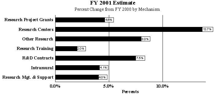
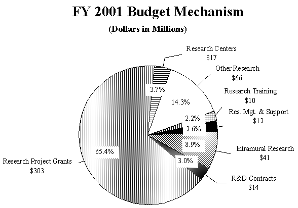
Promises for advancement in medical research are dependent on a continuing supply of new investigators with new ideas. In the Fiscal Year 2001 request, NEI will support 268 pre- and postdoctoral trainees in full-time training positions. Stipends will increase by 2.2 percent over Fiscal Year 2000 levels. The Fiscal Year 2001 request includes funding for 39 research centers, 209 other research grants, including one new clinical career award, and 35 R&D contracts. The mechanism distribution by dollars and percent change are displayed below:
1Sperduto R, Seigel D, Roberts J, Rowland M: Prevalence of myopia in the United States. Arch Ophthalmol 101(3):405-407, 1983.
2Steinberg EP, Javitt JC, Sharkey PD, et al: The content and cost of cataract surgery. Arch Ophthalmol 111:1041-1049, 1993.
3Ellwein LB, Friedlin V, McBean AM, Lee PP: Use of eye care services among the 1991 Medicare population. Ophthalmology 103(11):1732-12743, 1996.
4Rahmani B, Tielsch J, Katz J, et al: The cause-specific prevalence of visual impairment in an urban population, the Baltimore Eye Survey. Ophthalmology 103(11): 1721-1726, 1996.
5Kahn HA and Moorhead HB: Statistics on blindness in the model reporting area, 1969- 1970. U.S. Department of Health, Education, and Welfare. Public Health Service. Publication No. (NIH)73-427. pp. 120-143.
6Hillis A, et al: The evolving concept of amblyopia: a challenge to epidemiologists. Am J Epidemiol 118(2):192-205, 1983.
7Preslan M, Novak A: Baltimore Vision Screening Project. Ophthalmology 103(1):105- 109, 1996.
8Tielsch J, Sommer A, Will K, Katz J, Royall R: Blindness and visual impairment in an American Urban population, the Baltimore Eye Survey. Archives of Ophthalmology 108:286- 290, 1990.
June 2001