


Inside Structures of Life
Flash version (requires free Adobe Flash Player)
HTML Accessible Version
Slide 1
- Overview of structural biology
- Student snapshots
- Learning aids
- Glossary
- “Got It?” review questions
“… [offers] critical reading lessons to show students the real world applications of biology.” —High school teacher, Maryland
Slide 2

- Read about
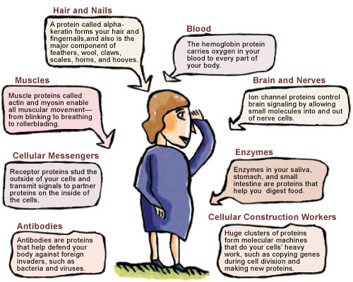
- The many roles of proteins
- Why protein shapes are important
- Tools used to study proteins
- How protein shapes affect the design of new medicines
Slide 3
Learn About
- How proteins keep us alive and healthy
- How faulty proteins can cause diseases
Slide 4
Learn About
- How protein shapes are being used to design new medicines
- How protein shapes are teaching us about biology
Slide 5
- Scientists
- Student Researchers


Slide 6
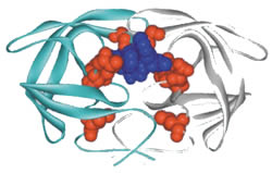
Tools for Studying Proteins
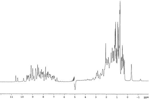
- X-ray crystallography
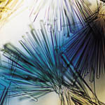

- Nuclear magnetic resonance (NMR) spectroscopy

Slide 7
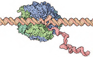
Computer Graphics Are Key

Slide 8
Read About
- The life cycle of the AIDS virus
- How the shape of a protein led to an AIDS drug

Slide 9