| Scientists Uncover New Clues About
Brain Function in Human Behavior
Researchers at the National Institute of Mental Health
(NIMH), part of the National Institutes of Health, have
discovered a genetically controlled brain mechanism
responsible for social behavior in humans — one
of the most important but least understood aspects of
human nature. The findings are reported in Nature
Neuroscience, published online on July 10, 2005.
The study compared the brains of healthy volunteers
to those with a genetic abnormality, Williams Syndrome,
a rare disorder that causes unique changes in social
behavior. This comparison enabled the researchers to
both define a brain circuit for social function in the
healthy human brain, and identify the specific way in
which it was affected by genetic changes in Williams
Syndrome.
People with Williams Syndrome who are missing about
21 genes on chromosome seven are highly social and empathetic,
even in situations that would elicit fear and anxiety
in healthy people. They will eagerly, and often impulsively,
engage in social interactions, even with strangers.
However, they experience increased anxiety that is non-social,
such as fear of spiders or heights (phobias) and worry
excessively.
For several years, scientists have suspected that abnormal
processing in the amygdala, an almond-shaped structure
deep in the brain, may be involved in this striking
pattern of behavior. The amygdala’s response and regulation
are thought to be critical to people’s social behavior
through the monitoring of daily life events such as
danger signals. Scientists know from animal studies
that damage to the amygdala impairs social functioning.
“Social interactions are central to human experience
and well-being, and are adversely affected in psychiatric
illness. This may be the first study to identify functional
disturbances in a brain pathway associated with abnormal
social behavior caused by a genetic disorder,” said
NIMH Director Thomas R. Insel, M.D.
In this study, investigators used functional brain
imaging (fMRI) to study the amygdala and structures
linked to it in 13 participants with Williams Syndrome
who were selected to have normal intelligence (Williams
Syndrome is usually associated with some degree of mental
retardation or learning impairment) and compared to
healthy controls. Andreas Meyer-Lindenberg, M.D., Ph.D.,
and Karen Berman, M.D., from the NIMH Genes, Cognition,
and Psychosis Program, and colleagues, then showed participants
pictures of angry or fearful faces. Such faces are known
to be highly socially relevant danger signals that strongly
activate the amygdala. The fMRI showed considerably
less activation of the amygdala in participants with
Williams Syndrome than in the healthy volunteers (see
graphic below). These findings suggest that reduced
danger signaling by the amygdala in response to social
stimuli might be responsible for their fearlessness
in social interactions.
Next, researchers showed the study participants pictures
of threatening scenes (a burning building or a plane
crash), which did not have any people or faces in them
and thus had no immediate social component. In remarkable
contrast to the response to faces, the amygdala response
to threatening scenes was abnormally increased in participants
with Williams Syndrome (see
graphic below), mirroring their severe non-social
anxiety.
“The amygdala response perfectly reflected the unique
profile of social and non-social anxiety in Williams
Syndrome,” said Meyer-Lindenberg. “Because our data
showed that the amygdala did still function, although
abnormally, in Williams Syndrome, we wondered whether
it might be its regulation by other brain regions that
was the cause of the amygdala abnormalities.”
To investigate this, the scientists looked at the whole
brain to identify other regions where reactivity was
different between Williams’s participants and healthy
volunteers. They identified three areas of the prefrontal
cortex, located in the front part of the brain, that
have been implicated in decision-making, representation
of social knowledge, and judgment. Those regions are
the dorsolateral, the medial, and the orbitofrontal
cortex. Specifically, the dorsolateral area is thought
to establish and maintain social goals governing an
interaction; the medial area has been associated with
empathy and regulation of negative emotion; and orbitofrontal
region is involved in assigning emotional values to
a situation.
The researchers found a delicate network by which these
three regions modulate amygdala activity. In Williams
Syndrome, this fragile system was significantly abnormal,
particularly the orbitofrontal cortex. This area did
not activate for either task and was not functionally
linked to the amygdala, as it was in healthy controls.
Instead, the scientists observed increased activity
and linkage in the medial region, which is consistent
with the high level of empathy exhibited by people with
Williams Syndrome.
“We had previously seen that the orbitofrontal cortex
is structurally abnormal in Williams Syndrome, but we
didn’t know what role it played functionally in the
disorder; it is now clear that this area can play a
major role in producing social behavioral abnormalities,” said
Berman. “The over-activity of the medial-prefrontal
cortex may be compensatory, but the result is still
an abnormal fear response. The medial-prefrontal cortex
still works and in fact it is working over-time because
it may be the only thing that still regulates the amygdala
in Williams Syndrome.”
Other releases on this topic: http://www.nimh.nih.gov/press/prwilliams.cfm.
For more information visit the NINDS web site on Williams http://www.ninds.nih.gov/disorders/williams/williams.htm.
In addition to the NIMH Intramural Research Program,
the research was also funded by a grant from the National
Institute on Neurological Disorders and Stroke (NINDS)
to co-author Dr. Carolyn Mervis, University of Louisville.
Also participating in the research were Dr. Ahmad Hariri,
Karen Munoz, Dr. Venkata Mattay, NIMH, and Dr. Colleen
Morris, University of Nevada.
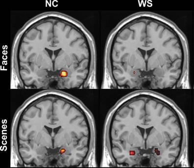 |
| Abnormal regulation
of the amygdala in participants with Williams
Syndrome (right) compared to controls (left).
The amygdala activates more for threatening
scenes (bottom), but less for threatening faces
(top). |
NIMH and NINDS are part of the National Institutes
of Health (NIH), the Federal Government's primary
agency for biomedical and behavioral research. NIH
is a component of the U.S. Department of Health and
Human Services.
The National Institutes of Health (NIH) — The
Nation's Medical Research Agency — is comprised
of 27 Institutes and Centers and is a component of
the U. S. Department of Health and Human Services.
It is the primary Federal agency for conducting and
supporting basic, clinical, and translational medical
research, and investigates the causes, treatments,
and cures for both common and rare diseases. For more
information about NIH and its programs, visit www.nih.gov. |