What Is a Coronary Calcium Scan?
A coronary calcium scan is a test that can help show
whether you have
coronary
artery disease (CAD). In CAD, a fatty material called plaque (plak) narrows
your coronary (heart) arteries and limits blood flow to your heart. CAD is the
most common type of heart disease in both men and women. It can lead to
angina,
heart
attack,
heart
failure, and
arrhythmia.
Coronary calcium scanning looks for specks of
calcium (called calcifications) in the walls of the coronary arteries.
Calcifications are an early sign of heart disease. The test can show, before
other signs and symptoms occur, whether you’re at increased risk for a
heart attack or other heart problems.
A coronary calcium scan is most useful for people
who are at moderate risk for a heart attack. You or your doctor can calculate
your 10-year risk using the
Risk
Assessment Tool from the National Cholesterol Education Program. People at
moderate risk have a 10 to 20 percent chance of having a heart attack within
the next 10 years. The coronary calcium scan may help doctors decide who within
this group needs treatment.
Two machines can show calcium in the coronary
arteries—electron beam computed tomography (EBCT) and multidetector
computed tomography (MDCT). Both use an x-ray machine to make detailed pictures
of your heart. Doctors study the pictures to see whether you’re at risk
for heart problems in the next 2 to 10 years.
A coronary calcium scan is simple and easy for the
patient, who lies quietly in the scanner machine for about 10 minutes. Pictures
of the heart are taken that show whether the coronary arteries have
calcifications. (For more information, see “What To Expect During a Coronary Calcium
Scan.”)
Coronary Calcium
Scan
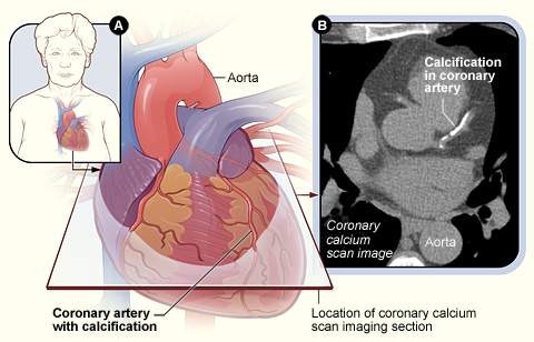
Figure A shows the position of the
heart in the body and the location and angle of the coronary calcium scan
image. Figure B is the coronary calcium scan image showing calcification in a
coronary artery.
April 2008
|