| |
| |
| Peter A. Bandettini, Ph.D., Investigator |
 |
Dr. Bandettini received his B.S. from Marquette University in 1989 and his Ph.D. from the Medical College of Wisconsin in 1994, where he pioneered the development of magnetic resonance imaging of human brain function using blood oxygenation contrast. During his postdoctoral fellowship at the Massachusetts General Hospital with Bruce Rosen, he continued his investigation of methods to increase the interpretability, resolution, and applicability of functional MRI techniques. In 1999, he joined NIMH as an Investigator in the Laboratory of Brain and Cognition and as the Director of the NIH Functional MRI core facility. In 2001, he was awarded the Scientific Director's Merit Award for his efforts in establishing the NIH FMRI core facility. In 2002, he was awarded the Wiley Young Investigator's at the annual Organization for Human Brain Mapping Meeting. His laboratory is currently developing MRI methods improve the spatial resolution, temporal resolution, sensitivity, interpretability, and applications of functional MRI.
|
|
Staff:
- Dr. Rasmus M. Birn, Ph.D., Staff Scientist, (301) 402-1350 rbirn@mail.nih.gov
- Dr. Anthony Boemio, Ph.D., Postdoctoral Fellow, (301) 402-1379 anthonyboemio@mail.nih.gov
- Dr. Hauke Heekeren, M.D., Ph.D., Adjunct Investigator hauke.heekeren@charite.de
- Dr. David Knight, Ph.D., Postdoctoral Fellow, (301) 402-1359 knightd@mail.nih.gov
- Dr. Nikolaus Kriegeskorte, Ph.D., Postdoctoral Fellow, (301) 594-9195 kriegeskorten@mail.nih.gov
- Ms. Kay Kuhns, B.S., Program Specialist, (301) 594-9191 kuhnsb@mail.nih.gov
- Dr. Marta Maieron, Ph.D., Adjunct Investigator mmaieron@makek.dstb.uniud.it
- Ms. Hanh Nguyen, B.S., Post baccalaureate Fellow, (301) 402-7298 htnguyen@niaid.nih.gov
- Ms. Natalia Petridou, M.S., Student petridon@mail.nih.gov
- Mr. Douglass Ruff, Post baccalaureate Fellow, (301) 451-9582 ruffd@mail.nih.gov
- Ms. Monica Smith, B.S., Post baccalaureate Fellow, (301) 594-9197 monicasmith@mail.nih.gov
- Mr. August Tuan, B.S., Adjunct Investigator august@salk.edu
- Ms. Najah Waters, B.S., Post baccalaureate Fellow, (301) 402-7299 nw92i@nih.gov
Research Interests:
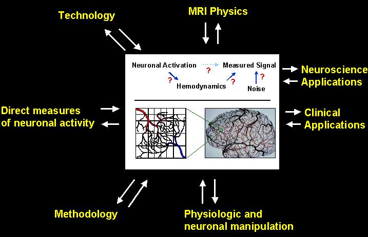
Functional MRI is a technique that utilizes time series collection of rapidly-obtained magnetic resonance images sensitive to brain activation induced changes in blood flow, oxygenation, and volume. The utility of Functional MRI (fMRI) has been improving since it�s inception in 1991. The limits of the technique reside in the imaging technology, methodology, and in the uncertainly and variability in cerebral hemodynamics and neurovascular coupling. To improve the depth, breadth, and sophistication of the questions being addressed and the comparisons being made, it is necessary to develop ways to better characterize the hemodynamic response and to determine how precisely it relates to neuronal activity. It is also necessary to develop the technology and methodology that utilizes this information for making more accurate and interpretable maps.
While progress is being made, fMRI is still not in common clinical use and is outside the domain of many critical neuroscience issues. A few of the limitations are as follows: Functional MRI cannot map transient activity on the order of milliseconds. It cannot map brain activity on a spatial scale smaller than about 2 mm2. Because of baseline drift, it cannot map very slow �state� changes. It cannot differentiate activity resulting from excitatory vs. inhibitory input. It cannot map baseline activity and metabolic state. It cannot temporally resolve cascaded communication between sequentially activated brain regions. Calibration procedures are relatively crude. It cannot draw inferences about individual activation maps as they relate to averaged population brain activation maps. Progress towards overcoming most if not all of these limitations can be made by a research strategy which is at the interface of applications and fundamental developments
The Unit on Functional Imaging Methods is a team of are a team of physicists, psychologists, engineers, neuroscientists, and computer scientists committed to developing fMRI to it�s full potential through interrelated advancements in technology, methodology, interpretation, and applications.
|
Selected Recent Publications:
R. M. Birn, R. W. Cox, P. A. Bandettini (2004) Functional MRI experimental designs and processing strategies for studying brain activation associated with overt responses., NeuroImage 23, 1046-1058.
P.S.F. Bellgowan, Z. S. Saad, P. A. Bandettini (2003) Understanding neural system dynamics through task modulation and measurement of functional MRI amplitude, latency, and width., Proc. Nat'l. Acad. Sci. USA 100, 1415-1419.
Z. S. Saad, K. M. Ropella, E. A. DeYoe, P. A. Bandettini (2003) The spatial extent of the BOLD response, NeuroImage 19, 132-144.
D. C. Knight, H. T. Nguyen, P. A. Bandettini (2003) Expression of conditional fear with and without awareness., Proc. Nat'l. Acad. Sci. USA 100, 15280-15283.
J. C. Patterson II, L. G. Ungerleider, and P. A Bandettini (2002) Task - independent functional brain activity correlation with skin conductance changes: an fMRI study, NeuroImage 17, 1787-1806.
J. Bodurka, P. A. Bandettini (2002) Toward direct mapping of neuronal activity: MRI detection of ultra weak transient magnetic field changes, Magn. Reson. Med. 47, 1052-1058.
R. M. Birn, R. W. Cox, P. A. Bandettini (2002) Detection versus estimation in event-related fMRI: choosing the optimal stimulus timing, NeuroImage 15, 262-264.
All Selected Publications
Contact Information:
Dr. Peter A. Bandettini
Functional Imaging Methods Unit
Laboratory of Brain and Cognition, NIMH
Building 10, room 1D80
10 Center Drive, MSC 1148
Bethesda, MD 20892-1148
Telephone: (301) 402-1333 (office),
(301) 402-1370 (fax)
Email: bandettini@nih.gov
|
|