| |

“The findings and conclusions in this review are those of the author(s) and do not
necessarily represent the views of the funding agency.” |
| This article was published with modifications in Epidemiologic Reviews 2000; 22(2):203-217 |
|
by Joseph M. Zmuda, Ph.D., 1 Jane A. Cauley, Dr.PH, 1 Robert E. Ferrell, Ph.D. 2
Authors’ affiliations:
From the Departments of Epidemiology1 and Human Genetics,2Graduate School of Public Health, University of Pittsburgh, Pittsburgh, PA
This work was supported in part by the United States Public Health Service grants 1P60 AR-44811 and P30 DK 46206.
| Address correspondence to: |
Joseph M. Zmuda, Ph.D.
Department of Epidemiology
Graduate School of Public Health
University of Pittsburgh
130 Desoto St.
Pittsburgh, PA 15261
Phone: 412-624-629
Fax: 412-624-7397
Email: EPIDJMZ@PITT.EDU |

|
ABSTRACT |
NAD(P)H:quinone oxidoreductase (NQO1) catalyzes the 2- or 4-electron reduction of numerous endogenous and environmental quinones (e.g. the vitamin E  -tocopherol quinone, menadione, benzene quinones). In laboratory animals treated with various environmental chemicals, inhibition of NQO1 Vitamin D regulates calcium homeostasis and bone mineralization, and vitamin D action is mediated through the vitamin D receptor (VDR), a ligand activated transcription factor. The gene encoding the VDR is located on chromosome 12q, and has several known allelic variants including a Fok I restriction fragment length polymorphism in exon II, BsmI and ApaI polymorphisms in the intron between exons VIII and IX, a synonymous Taq I variant in exon IX, and a mononucleotide [(A)n] repeat polymorphism in the 3' untranslated region. -tocopherol quinone, menadione, benzene quinones). In laboratory animals treated with various environmental chemicals, inhibition of NQO1 Vitamin D regulates calcium homeostasis and bone mineralization, and vitamin D action is mediated through the vitamin D receptor (VDR), a ligand activated transcription factor. The gene encoding the VDR is located on chromosome 12q, and has several known allelic variants including a Fok I restriction fragment length polymorphism in exon II, BsmI and ApaI polymorphisms in the intron between exons VIII and IX, a synonymous Taq I variant in exon IX, and a mononucleotide [(A)n] repeat polymorphism in the 3' untranslated region.
The individual allelic variants and their haplotypes have been widely studied as markers of susceptibility to osteoporosis, a prevalent metabolic bone disease characterized by reduced bone mass and a resultant increased susceptibility to fracture. Most reports have focused on the Bsm I site. The homozygous absence of the Bsm I site has been associated with a small to modest decrease in bone mass, and a 2-fold increase in the risk of hip fracture compared with the homozygous absence of this site in some studies. Attempts to replicate these findings, however, have yielded conflicting results. The strength of association has been modified by molecular variation in other genes and by other risk factors such as age and dietary calcium intake in some reports. This suggests that VDR allelic effects may be context dependent and that there may be larger VDR effects in certain subgroups in the population.
More recent studies suggest a possible role of VDR gene variants in the development of other diseases including breast and prostate cancer, osteoarthritis, atherosclerotic coronary artery disease, diabetes, primary hyperparathyroidism, infection, and psoriasis, although these findings require confirmation.Understanding the role of VDR gene variants in the susceptibility to osteoporosis and these other conditions may suggest novel approaches to their prevention and treatment.
Keywords: Vitamin D receptor, osteoporosis, bone mineral density, fracture, prostate cancer, breast cancer, osteoarthritis, hyperparathyroidism, diabetes, coronary artery disease, psoriasis, epidemiology.

|
GENE |
Genomic organization
The vitamin D receptor (VDR) belongs to the steroid and thyroid hormone receptor family of ligand activated transcription factors. The VDR mediates the effects of 1,25 dihydroxyvitamin D [1,25(OH) 2D] on gene expression (1). The gene encoding the VDR is located on chromosome 12cen-q12 (2), contains 14 exons (3), and spans approximately 75 kilobases of genomic DNA (4). Exons IA through IF encode the 5' untranslated region, exons II and III encode the DNA-binding domain, and exons IV-IX encode the ligand-binding region (3, 5).
|
GENE VARIANTS |
| At least 22 unique loss of function mutations in the VDR gene have been reported (6, 7). Single nucleotide changes producing amino acid substitutions in the DNA-and ligand-binding domains are the predominate type of mutation found (6). Less frequent mutations including premature stop codons, cryptic splice sites, and a partial gene deletion have also been described (6, 8, 9). These mutations cause hereditary vitamin D resistant rickets, a rare autosomal recessive disease resulting from target organ resistance to 1,25(OH)2D (6). An updated database of rare VDR mutations can be found on the Human Gene Mutation Database (http://www.uwcm.ac.uk/uwcm/mg/hgmd0.html  ). ).
Several common allelic variants have also been identified in the VDR gene and are the focus of the present review (Figure 1). The presence of a T/C transition polymorphism (ATG to ACG) at the first of two potential translation initiation sites in exon II (10) has been defined using the FokI restriction endonuclease (11). Individuals with the C allele (designated F) initiate translation at the second ATG site and lack the three NH2-terminal amino acids of the full-length VDR protein (12). In contrast, individuals with the T allele (designated f) initiate translation at the first ATG site and synthesize the full-length (427 amino acids) VDR protein (12). The ff genotype frequency was 4% among African-Americans and 13-18% among Asians and Caucasians in one report (13).
BsmI (14) and ApaI (15) restriction site polymorphisms occur in the intron separating exons VIII and IX (Figure 1). A T/C nucleotide substitution (ATT to ATC) leading to a synonymous change at codon 352 (isoleucine) in exon IX has also been described (16), and is detected by the restriction enzyme TaqI . The BsmI and FokI alleles do not appear to be in linkage disequilibrium (11, 13, 17, 18), whereas a strong concordance exists between the absence of the BsmI (B allele) and presence of the TaqI (t allele) sites (19), and these sites show significant linkage disequilibrium with the ApaI polymorphism. Hustmyer et al.(16) detected a rare third allele by ApaI digestion in African-Americans, but more recent PCR based typing of the ApaI polymorphism has not detected the presence of this allele. Morrison et al.(14) reported a fifth restriction site polymorphism, detected by southern blot analysis of EcoRV digested genomic DNA probed with a VDR cDNA probe, and Hustmyer et al.(16) showed that the frequency of the two alleles at this locus varied among Caucasians, African-Americans, and Asians. The molecular basis of this polymorphism is unknown, and recent studies using PCR based assays have not genotyped this variation. Because few studies of the EcoRV polymorphism are available, we have not reviewed this variant.
The BsmI bb genotype frequency was 2% among Asians, 5% among African-Americans, and 17% among Caucasians in a meta-analysis (20). The frequency of TaqI genotypes in these populations is similar to BsmI genotype frequencies. The ApaI AA genotype frequency is 9% among Asians (21), 28% among Caucasians (22), and 44% among African-Americans (23).
A mononucleotide repeat [(A)n] polymorphism which varies in length from 13-24 adenosines (12 alleles) [poly(A)] occurs in the 3' untranslated (UTR) region of the VDR gene (24). The distribution of allele size is bimodal, such that individuals can be classified as having short (A13-A17) or long (A18-A24) alleles (24). The frequency of short alleles in one study was 5-10% among Asians, 32% among African-Americans, and 41% among Caucasians (24). The longest alleles (A23-A24) in that study were only found among African-Americans, whereas the shortest allele (A13) was only found among Hispanics (24).
Linkage disequilibrium has been reported between the poly(A) and BsmI alleles, such that the short poly(A) and BsmI B alleles (BS haplotype) and the long poly(A) and BsmI b alleles (bL haplotype) are coupled. Linkage disequilibrium is nearly complete among Caucasian- (disequilibrium coefficient, 0.96) and Japanese-Americans (0.90), but is less pronounced among African-Americans (0.53) (24). Poly(A) and FokI alleles do not appear to be in linkage disequilibrium (25, 26).
Functional consequences of VDR gene variants
The possible functional consequences of VDR alleles remain unclear. The ApaI and BsmI variants are unlikely to have functional consequences since both sites are located in the intron between exons VIII and IX and neither variant is near the intron-exon boundaries or known to produce splicing errors. Moreover, several studies have found similar VDR protein (27-29) and mRNA levels (28, 30), ligand binding affinity (28), DNA binding (28), and transactivation function (28) between BsmI genotypes, although these observations have not been universal (31). The TaqI polymorphism is also unlikely to directly affect VDR function since both alleles code for isoleucine at amino acid 352.
Several studies have also examined the association between the common BsmI/ApaI/TaqI haplotypes and VDR function. Morrison et al.(19) showed that COS-7 and rat osteosarcoma cells transfected with reporter gene constructs containing the the baT haplotype had significantly lower luciferase activity than those with the BAt haplotype. Consistent with these observations, the baT haplotype has been associated with significantly lower VDR mRNA levels in parathyroid adenomas of patients with primary hyperparathyroidism (31). In contrast to these studies, Beaumont et al.(32) recently demonstrated significantly greater luciferase activity with reporter gene constructs containing the baT haplotype in transfected human osteoblast and osteosarcoma cell lines. One possible explanation for these inconsistent findings may be that the effect of VDR allelic variants on VDR function is tissue and/or species specific.
The FokI variant remains a candidate functional polymorphism. Colin et al. (33) found that phytohemoglutinin (PHA)-stimulated growth of peripheral blood monocytes differs by FokI genotype. They demonstrated that the ½ maximal concentration for 1,25(OH)2 vitamin D inhibition of PHA stimulated growth was significantly higher for cells containing the full-length VDR isoform (i.e., Ff and ff genotypes) than those with the shorter isoform (FF genotype). Interestingly, there were no genotype related differences in maximal inhibition of growth, raising the possibility that genotypic effects may be most apparent among individuals with low 1,25(OH)2 vitamin D levels. Transfection experiments in COS-7, HeLa, and fibroblast cell lines have also shown that the full-length VDR isoform has a decreased ability to induce transcriptional activation of reporter genes in response to 1,25(OH)2D compared with the shorter F allele isoform (12, 25), although these observations were not confirmed in another study (34). The f allele isoform interacts with the basal transcription factor IIB less efficiently than the F allele isoform, providing a possible mechanism for the reduced transactivation associated with this allele (35). The 3' poly(A) allelic variants do not appear to alter VDR mRNA stability (36).
|
DISEASE |
Osteoporosis
Osteoporosis is a prevalent metabolic bone disease characterized by low bone mass and a resultant increased susceptibility to fracture. The risk of fracture increases by as much as 2.5- to 3.0-fold with each standard deviation (SD) reduction in bone mass (37). Bone mass and osteoporotic risk are under strong genetic control (38), and the VDR gene has been widely studied as an osteoporosis candidate gene.
Most reports have examined the association between the BsmI polymorphism and bone mass. An initial study by Morrison et al. (19, 20, 39) documented about 0.5 SD or 8% lower (p<0.01) spine bone mass in a sample of pre- and postmenopausal Australian women with the BB compared with bb genotypes. These findings were confirmed in some but not all subsequent studies (38). In a meta-analysis of 16 reports published through July, 1996 and involving over 3,600 subjects, the BB genotype was associated with 0.2 SD or 2.4% lower hip (p=0.03) and 0.2 SD or 2.5% lower spine (p=0.06) bone mass compared with the bb genotype (20). More recently, Gong et al.(40) performed a qualitative meta-analysis of 75 reports and abstracts published up to January, 1997 and involving more than 14,000 individuals. They concluded that VDR alleles (B, A, t) were associated with lower hip and spine bone mass more often than the expected 5% false positive rate under the null hypothesis (40). Studies were more likely to find a significant association between VDR alleles and bone mass among premenopausal than postmenopausal women or pre- and postmenopausal women combined, and less likely to find a significant association if they included osteoporotic subjects. This suggests that the major effect of VDR genotype may be on peak bone mass, rather than on age- or menopause related bone loss.
Bone mass in the elderly is a product of both peak skeletal mass achieved during the first three decades of life, and subsequent age- and menopause-related rates of bone loss. Although less well studied, allelic variants of the VDR gene have not been consistently associated with rates of bone loss among postmenopausal women. Three (41-43) out of 8 studies (22, 23, 44-46) have found significantly greater postmenopausal bone loss associated with the B allele. Three studies did not find a significant association between the TaqI polymorphism and bone loss (22, 45, 47), although we documented a significantly greater rate of hip bone loss among older (>70 yr), but not younger (<70 yr), African-American women with the tt genotype (23). Most studies had fewer than 100 subjects and less than 2 years of follow-up, and may have lacked adequate statistical power to detect differences between genotypes. For example, a more than 2-fold greater rate of spinal bone loss among postmenopausal women (n=109) with the BB or tt genotype did not achieve statistical significance in one study (22). Postmenopausal Mexican American women with the FokI ff genotype experienced significantly greater hip bone loss compared to women with either the Ff or FF genotypes (11), although this finding was not confirmed in a subsequent study of Caucasian American women (48).
Two (49, 50) out of eight studies (51-56) have demonstrated a significant difference in VDR genotype or haplotype distribution between osteoporotic patients and controls. Most studies included fewer than 100 cases, so it is possible that small differences in genotype frequencies were missed. The largest study to date (50) found that the homozygous BAt haplotype was significantly more prevalent among 176 osteoporotic Italian women compared with 144 controls (24% vs 8%, respectively; p<0.01), whereas the homozygous baT haplotype was less common among osteoporotic women (7% vs 18%, respectively; p<0.01).
More recent studies have focused on the FokI variant in exon 2. Initial reports of this polymorphism found 11-12% (~1 SD) lower bone mass at the hip and spine in Japanese (12), Mexican-American (11), and Caucasian-American (57) women with the ff compared with FF genotypes. Subsequent reports have not confirmed significant associations between the FokI variant and bone mass, and differences between homozygous genotypes have generally been much smaller (~2-5% or <0.3 SD) (13, 17, 18, 48, 58-61). Most studies have lacked sufficient statistical power to detect differences of this magnitude. Moreover, ethnic (genetic) background may modify the effects of this polymorphism (57). There may also be effect modification by unlinked loci (modifier genes) (62) and environmental factors such as dietary calcium intake (43, 63-65) that remain to be fully explored.
The physiologic mechanisms mediating the associations between VDR gene variants and bone mass and bone loss are unclear, but are likely due to established actions of vitamin D on calcium homeostasis. For example, 1,25(OH)2D and its receptor mediates active intestinal calcium absorption (66), and calcium absorption has been reduced in subjects with the BB genotype (23, 67, 68) and homozygous BAt haplotype (68, 69). These associations may be more pronounced among subjects with low dietary calcium intake (67). Premenopausal women with the BAt haplotype had 11% lower (69) and postmenopausal women 37% lower (68) calcium absorption compared to women with the baT haplotype (p<0.05). Thus, the effect of VDR gene variation on calcium absorption may also be modified by age or hormonal status. An additive effect of FokI alleles on calcium absorption has also been demonstrated among children (70). Calcium absorption was 41.5% greater in children who were FF than ff homozygotes, and 17% greater in herterozygotes (70). However, associations between VDR genotype and calcium absorption have not been confirmed in all studies (27, 71, 72). Nevertheless, these results suggest that there may be VDR genotype dependent differences in intestinal sensitivity to 1,25(OH)2D.
Osteoporotic fracture
There have been relatively few studies of VDR gene variants and the risk of osteoporotic fractures (Table 1). An ecological analysis of 14 published studies suggested that higher population frequencies of the TT genotype are associated with lower age-adjusted hip fracture rates (78), consistent with studies showing that this polymorphism is associated with greater bone mass. Feskanich et al.(73) found a 2.4-fold greater risk (95% CI: 1.1, 5.2) of hip fracture associated with the BB compared with the Bb or bb genotypes in a nested case-control study of Caucasian-American women aged 43-69 yrs. The increased risk of fracture associated with the BB genotype in this study is much greater than that expected based on the small differences in bone mass associated with this polymorphism. Uitterlinden et al.(74) documented a relationship between the number of baT haplotypes and the risk of spine and non-spine fractures in a nested case-control study of older Caucasian European women. The risk of both fractures was 80% greater (95% CI: 1.0, 3.3) among heterozygous women, and 2.6-fold greater (95% CI: 1.4, 5.0) among homozygous women. The direction of the association conflicts with that found by Feskanich et al. (73), and suggests that different alleles may be associated with fracture in different populations (i.e., allelic heterogeneity). Interestingly, the increased risk of fracture associated with the baT haplotype was independent of bone mass, raising the possibility that factors other than low bone mass explain the association between VDR haplotype and fracture risk (74) Nevertheless, in the largest study to date, we were unable to confirm a relationship between the TaqI and ApaI variants, either alone or combined, and the incidence of hip, spine or other fractures in a case-cohort study nested within a prospective study of 9,704 Caucasian-American women aged 65 yrs and older (75). Analyses stratified by age (<75 vs >75 yrs), calcaneal bone mass (<0.40 vs >0.40 mg/cm2), and dietary calcium intake (<640 vs >640 mg/day) produced similar results.
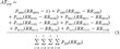 |
Table 1
Summary of Studies Examining the Association between VDR Genotype or Haplotype and Osteoporotic Fracture. |
The relationship between the FokI variant and fracture risk has been less well studied. Gennari et al. (61) found that the ff genotype was over-represented among postmenopausal women with vertebral fractures (25%) compared with controls (11%), equivalent to an odds ratio of 2.6 (95% CI: 1.4, 4.9). These findings have not been replicated in other populations yet.
Summary
Bone mass is under strong genetic control, but the specific genes and allelic variants contributing to bone mass and osteoporotic risk are not well defined (38). The VDR gene has been widely studied as an osteoporosis candidate gene during the past several years, with most reports focusing on a BsmI restriction fragment length polymorphism in intron 8. The homozygous absence of this site has been associated with a small decrease (2%) in bone mass in a large meta-analysis, and with an increase in hip fracture risk in one study, although attempts to replicate these later findings have been unsuccessful. A potentially functional FokI polymorphism in exon 2 has also been associated with modest differences in bone mass in some studies and with vertebral fracture risk in one report, although again these findings have been inconsistent.
Conflicting results are not unexpected in association studies and may arise for several reasons including differences in ethnic (genetic) background, gene-gene and gene-environment interactions, and the definition of the phenotype. Inappropriate selection of controls is the major confounding factor in association studies, however, and differences in subject ascertainment may also contribute to discrepant and sometimes spurious results. For instance, the distribution of VDR genotypes was not in Hardy-Weinberg equilibrium (i.e., genotype frequencies were not predicted by allele frequencies) in some studies. Departures from Hardy-Weinberg equilibrium may arise for several reasons apart from genotyping errors including chance fluctuations due to small samples, nonrandom mating, migration into or out of the population, selective survivorship among genotypes, population stratification, and admixture of different ethnic groups (79). Deviations from Hardy-Weinberg equilibrium can bias the type I error rate such that the chance of a false-positive association increases substantially if the proportion of homozygotes with the high-risk allele is more common in the general population than predicted by Hardy-Weinberg equilibrium (80). Appropriate selection of controls is thus essential in association studies, but can be difficult due to unrecognized confounding by ethnic, ancestry, or admixture differences between cases and controls. Family-based association tests, such as the transmission-disequilibrium test (TDT), avoid confounding due to population stratification or admixture (81-83), but have rarely been used in studies of VDR alleles (84). The TDT test compares allele frequencies in cases with the frequencies of nontransmitted alleles in parents, thereby eliminating the need for ethnically matched controls. Recent modifications to the TDT make it a more practical tool for the study of quantitative traits such as bone mass (85). Future investigations of VDR gene variation should use family-based association methods to validate the results of population-based studies.
|
ASSOCIATIONS |
Other Diseases
Cancer
Vitamin D can inhibit cancer cell growth, angiogenesis and metastasis (86), and recent reports suggest that common VDR gene variants may be associated with the risk of prostate and breast cancer. At least 10 published reports have examined the relationship between VDR allelic variants and prostate cancer (Table 2). Initial reports found 70-80% lower risk of prostate cancer associated with the TaqI tt genotype (87) or short poly(A) alleles (88). Subsequent studies have been inconsistent, and generally not confirmed an association between these polymorphisms and the overall risk of prostate cancer (89-91, 94, 96). However, associations were stronger for more advanced disease in some reports (88, 89), suggesting that VDR allelic variants may influence the progression, rather than initiation, of prostate cancer.
| |
Table 2
Summary of Studies Examining the
Association between VDR Genotype and Prostate Cancer. |
Vitamin D may also play a role in normal prostate growth (97), and one recent study demonstrated an association between the VDR BsmI polymorphism and risk of benign prostatic hypertrophy (BPH) (95). Thus, inclusion of men with BPH as controls may have masked or attenuated an association between VDR polymorphisms and prostate cancer in some studies.
At least 7 studies have examined the association between VDR allelic variants and breast cancer risk (Table 3). An initial report found nearly 4-fold greater risk of breast cancer associated with the homozygous presence of the BsmI site among Japanese women (103) which is consistent with the 3-fold increases in prostate cancer risk among Japanese men with this VDR genotype (95). Subsequent reports have demonstrated similar (>2-fold) increases in breast cancer risk among women homozygous for the presence of the ApaI (99), FokI (26), or short poly(A) alleles (98), although these findings have not been universal (98, 99) and in one study the homozygous presence of the BsmI site was associated with a decreased risk of breast cancer among Latina women (98). In two studies, an association was found for VDR genotype and metastatic, but not overall, disease risk (101, 102), suggesting that VDR allelic variants may influence tumor progression rather than development.
| |
Table 3
Summary
of Studies Examining the Association between VDR Genotype and Breast Cancer. |
Osteoarthritis
Vitamin D receptor allelic variants have also been associated with prevalent osteoarthritis (OA) in some studies. The presence of the baT haplotype (104) or T allele (105) was associated with an approximately 2.5-fold increase in the risk of knee OA, which was independent of age, body mass index, and bone mass in two case-control studies. This relationship was largely explained by an association with osteophytes rather than joint space narrowing in one study (104), suggesting that VDR genotype may influence particular features of OA. Biological support for this association comes from studies showing that serum levels of vitamin D are related to the progression of knee OA (106), and that vitamin D receptors are expressed in chondrocytes (107), a cellular component of osteophytes (108). In contrast to these findings, the TaqI T allele was associated with a decreased risk of spine OA (109), and the BsmI variant was not significantly associated with hip OA (total hip replacement) (110) in other studies. These studies are limited by their cross-sectional design, small sample size, and focus on Caucasian subjects. Prospective studies in larger and more diverse populations are needed to test whether VDR genotypes and haplotypes are associated with the incidence and progression of radiographically defined OA.
Hyperparathyroidism
The vitamin D receptor mediates the inhibitory effects of vitamin D on parathyroid hormone (PTH) secretion (111) and parathyroid cell proliferation (112, 113). Recent studies suggest that VDR gene variants may be associated with primary hyperparathyroidism (31, 114-117), a common disease often caused by benign parathyroid adenoma or parathyroid hyperplasia and accompanied by excessive PTH secretion (118). Carling et al. (114, 116) found that the b, a and T alleles were significantly more common among patients with primary hyperparathyroidism than among age-matched controls. The estimated risk of primary hyperparathyroidism was 2.5-fold greater (95% CI: 1.3, 5.1) among women with the baT haplotype compared to those without this haplotype (116). Consistent with these findings, PTH mRNA levels were nearly 60% higher among patients with the baT haplotype compared to those with other haplotypes (31). The presence of the BsmI (119,120) and ApaI (121) restriction sites has also been associated with elevated PTH levels in patients with end-stage renal disease, suggesting that VDR gene variants may influence the development or severity of secondary hyperparathyroidism in such patients.
Diabetes
Transmission disequilibrium testing in 93 Indian families revealed that the b allele and bT and bAT haplotypes are preferentially transmitted from parents to offspring affected with Type I diabetes (84). Insulin secretion was 30-50% lower (p<0.05) in non-diabetic Bangladeshi Asians with the bb, aa or TT genotypes compared with the BB, AA or tt genotypes, respectively (122). These results are consistent with the presence of vitamin D receptors in pancreatic b-cells (123), and with studies showing that vitamin D deficiency impairs insulin secretion (124) and that vitamin D treatment prevents the development of Type I diabetes in the nonobese diabetic mouse model (125).
Coronary artery disease
The risk of prevalent ECG-confirmed myocardial infarction increased by 20% (95% CI: 1.0, 1.5) per copy of the baT haplotype in a population-based study of men and women aged 55-80 years (126). This association was independent of traditional risk factors for myocardial infarction including age, obesity and serum levels of total and high-density lipoprotein cholesterol. Consistent with these findings, patients (n=41) undergoing open-heart surgery with the bb genotype were 4-times more likely (95% CI: 0.8, 22.5; p=0.09) to have severe coronary artery stenosis compared to those with the Bb or BB genotypes (127). Biological support for these associations comes from studies demonstrating that vitamin D receptors are present in aortic endothelial (128) and vascular smooth muscle (129) cells.
Infectious Diseases
The immune system is a well-known target of vitamin D (130), and children with hereditary vitamin D resistant rickets may have impaired phagocytosis and neutrophil motility, and an increased number and severity of infections (131). Moreover, administration of 1,25(OH)2D inhibits growth of Mycobacterium tuberculosis in human macrophages and monocytic cells in vitro (132). Bellamy et al. (133) reported that the TaqI tt genotype was significantly under-represented in patients infected with pulmonary tuberculosis (6.6%) and hepatitis B (7.3%) compared with controls (12% and 14%, respectively). A smaller, subsequent study also noted a lower frequency of the tt genotype among tuberculosis patients (6%) compared with their uninfected contacts (11%), although this difference did not achieve statistical significance (p=0.49) (134). However, there was significant interaction between 25-hydroxycholecalciferol status and VDR genotype (134). The combination of the TT/Tt genotype and 25- hydroxycholecalciferol deficiency was associated with a 2.8-fold (95% CI: 1.2, 6.5) increased risk of tuberculosis. A similar interaction between the FokI ff genotype and vitamin D status was also observed. Roy et al.(135) also found that the TaqI polymorphism is associated with susceptibility to Mycobacterium leprae infection in general and also to leprosy type. The estimated risk of tuberculoid leprosy was 3-fold greater (95% CI: 1.5, 7.1) among Bengali subjects with the tt compared with TT genotypes. In contrast, there was a 67% increase (95% CI: 1.02, 2.75) in the risk of lepromatous leprosy in subjects with the TT compared with tt genotypes. The possibility that common molecular variation in the VDR gene makes a broader contribution to host susceptibility to infectious diseases merits further investigation.
Psoriasis
Psoriasis is a chronic skin disease characterized by hyperproliferation of keratinocytes and inflammation (136). The observations that keratinocytes contain receptors for 1,25(OH)2D (137), and that active metabolites of vitamin D inhibit proliferation of these cells (138), prompted recent studies of the association between VDR allelic variants and psoriasis (139-141). The frequency of ApaI A allele was significantly more common among 104 psoriatic Korean patients (0.317) compared with 104 controls (0.168), equivalent to a 2.4-fold (95% CI: 1.3, 4.3) increase in disease risk among subjects with the Aa genotype and 5.0-fold (95% CI: 1.3, 19.1) increase in risk among those with the AA genotype (139). The age of onset of psoriasis was 19.1 yrs in patients with the AA genotype compared to 21.5 yrs in heterozygous subjects and 29.3 yrs in those with the aa genotype (p<0.05). However, Mee et al.(142) did not demonstrate an association between the BsmI polymorphism and psoriasis (175 cases) or response to calcipotriol in 92 patients with chronic psoriasis. Likewise, Kontula et al. (141) were unable to document a difference in BsmI allele and genotype distribution between psoriatic patients who did (n=10) and did not (n=9) respond to topical calcipotriol treatment.
|
INTERACTIONS |
The risk of osteoporosis associated with VDR genotype may be modified by age, diet, and other lifestyle factors. Failing to account for such interactions may mask an association with VDR genotype. For instance, an increased risk of hip fracture associated with the BB genotype was greatest among women who were older, leaner, and less active, and among those with lower dietary calcium intake in one study (73). Other small clinical trials found that VDR genotype is associated with the bone mass response to vitamin D supplementation (143, 144). Two exercise intervention studies (145, 146) did not find significant VDR genotype differences in changes in bone mass, perhaps because of their small sample size (< 35 subjects). Larger trials will be necessary to convincingly demonstrate that VDR genotype influences the response to dietary or lifestyle modifications. Nevertheless, research addressing the influence of gene-environment interactions may suggest novel strategies for preventing or delaying the onset of this disease.
The association between VDR gene variation and risk of osteoporosis may also be modified by allelic variation in other candidate genes. For example, Willing et al.(62) found that the BsmI polymorphism alone was not significantly associated with bone mass at the spine among premenopausal Caucasian women. However, bone mass was 15% or more than one SD lower among women with the BB genotype who were also homozygous for the absence of a PvuII variant in intron one of the estrogen receptor alpha (ESR1) gene (p<0.05 for interaction). In another report, a complex interaction between the two-locus VDR-ESR1 genotype and hormone replacement therapy in modifying calcaneal ultrasound measures was documented (147). These interactions are biologically plausible since estrogen can increase the number and expression of VDR in osteoblasts (148, 149) and duodenal mucosa cells (150). The risk of fracture per copy of the baT haplotype was 1.1 (0.7, 1.6) among women with the G/G genotype at an Sp1 binding site in the type Ia1 collagen gene, and 2.6 (1.6, 4.5) among those with the G/T or T/T genotype (p<0.05 for interaction) (74). Thus, the influence of VDR genotype on osteoporotic risk may depend on the presence or absence of allelic variants at other unlinked loci.
The effect of VDR polymorphisms on the risk of other diseases may also be context dependent, although few studies to date have examined possible interactions between VDR polymorphisms and environmental exposures. The Physicians Health Study, for example, found a significant reduction in prostate cancer risk associated with the VDR BB or tt genotypes, but only among men with the lowest serum 25(OH) vitamin D levels (90). Thus, future investigations of VDR genotypes and the risk of other diseases may need to assess and stratify by serum vitamin D levels. It will also be important to test for possible interactions between VDR alleles and molecular variation in other candidate genes.
|
LABORATORY TESTING |
Standard polymerase chain reaction and restriction fragment length polymorphism analysis has been widely used to detect the most common VDR gene variants (BsmI, ApaI, TaqI, FokI). A method using gradient polyacrylamide gel and silver staining techniques to directly haplotype the BsmI, ApaI, and TaqI restriction sites has also been described (151). The poly(A) variant is determined by polymerase chain reaction amplification of the 3' untranslated region using one [g33P]adenosine triphosphate end-labeled primer and one unlabeled primer (88). The products are then separated on 6% polyacrylamide sequencing gels and autoradiographed.
|
POPULATION TESTING |
| There is currently insufficient evidence implicating VDR gene variants in the etiology of osteoporosis and other diseases to justify population testing.
|
TABLES AND FIGURES |
Tables
Tables for allele/genotype frequencies
Figure
|
REFERENCES |
List of References
|
INTERNET SITES |
General Resources
Genetic Databases
|
|
|
Provides link to non-governmental sites and does not necessarily represent the views of the Centers for Disease Control and Prevention. |
|
|
Page last reviewed: June 8, 2007 (archived document)
Page last updated: November 2, 2007
Content Source: National Office of Public Health Genomics |
|