ISSN: 1080-6059
Volume 13, Number 5–May 2007
Dispatch
Gulf Coast Ticks (Amblyomma maculatum) and Rickettsia parkeri, United States
John W. Sumner,* Lance A. Durden,† Jerome Goddard,‡ Ellen Y.
Stromdahl,§ Kerry L. Clark,¶ Will K. Reeves,* and Christopher D. Paddock* ![]()
*Centers for Disease Control and Prevention, Atlanta, GA,
USA; †Georgia Southern University, Statesboro, GA, USA; ‡Mississippi Department
of Health, Jackson, Mississippi, USA; §US Army Center for Health Promotion and
Preventive Medicine, Aberdeen Proving Ground, Maryland, USA; and ¶University of
North Florida, Jacksonville, Florida, USA
Suggested citation for this article
Abstract
Geographic distribution of Rickettsia parkeri in its
US tick vector, Amblyomma maculatum, was evaluated by PCR. R. parkeri was detected in ticks from Florida, Georgia, Kentucky, Mississippi, Oklahoma,
and South Carolina, which suggests that A. maculatum may be responsible
for additional cases of R. parkeri rickettsiosis throughout much of its US
range.
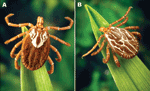 |
Figure. Adult Amblyomma maculatum (the Gulf Coast tick). A) Female; B) Male. Photographs courtesy of James Gathany, Centers for Disease Control and Prevention. |
The Gulf Coast tick, Amblyomma maculatum (Figure), is a Nearctic and Neotropical hard tick found in coastal areas of the southern United States, with inland range extensions in Kansas, Oklahoma, and some other states. It is also found in regions of several Central and South American countries that border the Gulf of Mexico and Caribbean Sea, including Mexico, Guatemala, Belize, Nicaragua, Honduras, Costa Rica, Colombia, Venezuela, and some parts of Ecuador and Peru (1).
Rickettsia parkeri, a member of the spotted fever group rickettsiae, was initially identified in Gulf Coast ticks in 1937 (2). In 2004, the first confirmed human infection with R. parkeri was reported (3). Since that report, confirmed cases of R. parkeri rickettsiosis have been identified in other persons in Mississippi, Virginia, and possibly other US states (4–6). Only a few studies, each conducted >50 years ago, document the occurrence of R. parkeri in A. maculatum ticks (2,7,8). No contemporary surveys have documented the range of R. parkeri in the United States or the frequency of R. parkeri infection in collections of individual Gulf Coast ticks.
The Study
A. maculatum ticks collected during 1996–2005 were evaluated by molecular methods for evidence of infection with R. parkeri. Ticks were collected from various locations in Florida, Georgia, Kentucky, Mississippi, Oklahoma, and South Carolina. Most were questing adults collected from vegetation by using flannel cloth flags; a few crawling, nonattached, nonengorged adults were obtained (4 from a coyote and 3 from human hosts), and 1 engorged nymph was removed from a cotton rat. Ticks were preserved in 70% ethanol or frozen at –80°C until evaluation.
Most individual ticks were minced with a sterile scalpel blade, and DNA was extracted by using a QIAamp Mini Kit (QIAGEN, Inc., Valencia, CA, USA). Others were minced or crushed after freezing in liquid nitrogen, and DNA was extracted by using an IsoQuick nucleic acid extraction kit (ORCA Research, Bothell, WA, USA). DNA extracts were evaluated by using nested or heminested PCR assays designed to amplify a segment of the rompA gene. For the primary stage of each assay, 5 μL of extract and primers 190–70 and 190–701 (9) were used. For the nested reaction, 2 μL of completed primary reaction was used as template with primers 190-FN1 (5´-AAG CAA TAC AAC AAG GTC-3´) and 190-RN1 (5´-TGA CAG TTA TTA TAC CTC-3´); for the heminested reaction, primers 190–FN1 and 190–701 were used. All reactions were prepared by using a High Fidelity PCR Master Kit (Roche Diagnostics, Indianapolis, IN, USA) with final primer concentrations of 300 nmol in a total volume of 50 μL. Thermalcycler parameters for the primary stage consisted of an initial denaturation period of 2 min at 94°C, followed by 40 cycles of 15 s at 94°C, 30 s at 60°C, 45 s at 72°C, and a 5-min extension period at 72°C. For the nested and heminested stages, the annealing temperature was changed to 55°C and the number of cycles was reduced to 30.
PCR products (10 μL) were separated by electrophoresis in 2% agarose gels containing ethidium bromide. For each positive reaction, the remaining 40 μL was subjected to gel electrophoresis, and products of the appropriate size were excised. DNA was purified from the gel fragments by using the QIAquick Gel Extraction Kit (QIAGEN). Purified PCR products were sequenced using the PCR primers and the GenomeLab DTCS Quick Start Kit (Beckman Coulter, Fullerton, CA, USA). For some products, additional sequencing primers 190-SF3 (5´-GGT ACT ACT CCC GTA GGT C-3´) and 190-SR2 (5´-CCG GCA GTA AKA GTA ACA G-3´) were used to obtain complete sequences for both strands. Sequences were detected by using a Beckman CEQ 8000 automated sequencer. Sequence similarities were determined by using the BLAST program (version 2.0, National Center for Biotechnology Information, www.ncbi.nlm.nih.gov/blast). Sequence-length reaction products (excluding primers) were 590 bp for primary, 559 bp for heminested, and 540 bp for nested.
DNA of R. parkeri was amplified from 21 (11.5%) of 182 male and female adult A. maculatum ticks collected in Georgia (11/64), Florida (7/89), Kentucky (1/1), Mississippi (1/24), and South Carolina (1/4) and from 1 engorged nymph collected in Oklahoma (Table). A unique rompA sequence (GenBank accession no. EF372578) amplified from 5 adult female ticks collected in Florida, Georgia, and Mississippi showed closest homology (≈94%) to several other rompA sequences (GenBank accession nos. EF063690, AY093696, AF120021, and DQ365801). PCR amplification of a 208-bp segment of the rickettsial 17-kDa antigen gene (10) from these same 5 ticks (GenBank accession no. EF372579) showed 100% homology with the corresponding sequences of Rickettsia sp. Arahna (AY360215), R. montanensis (DQ402377), Rickettsia sp. Hf332 (AB114804), Rickettsia sp. Is-1 (DQ344620), and "R. gravesii" (DQ269436).
Precise estimates of infection prevalence could not be assessed from these data because most of the ticks evaluated in this study were collected as relatively small sample sizes or were collected in a discontinuous manner as multiple samples from the same sites over several weeks or months in a given year. However, some collections were flagged synchronously at a single location, including those in Copiah County, Mississippi, during July 2002 (n = 9) and in Franklin County, Florida, during July 2004 (n = 25) and July 2005 (n = 27). Infection prevalence for each of these 3 collections was 11%–12%, which suggests that R. parkeri may be a relatively common inhabitant of some populations of Gulf Coast ticks. By comparison, the estimated prevalence of infection of tick vectors with R. rickettsii, the etiologic agent of Rocky Mountain spotted fever (RMSF), is typically much lower, as determined by surveys elsewhere, which identified R. rickettsii in only 0.05%–1.3% of the collected specimens: 3,705 Dermacentor andersoni ticks from canyons bordering the Bitterroot Valley of Montana; 2,123 and 310 D. variabilis ticks from RMSF-endemic areas of North Carolina and Ohio, respectively; and 669 A. aureolatum ticks from São Paulo, Brazil (11–14).
Conclusions
R. parkeri has been isolated in culture from Gulf Coast ticks collected in Alabama, Florida, Georgia, Mississippi, and Texas ([2,7,8], C. Paddock, unpub. data). These results, combined with data from the present study, suggest that in the United States R. parkeri can be found anywhere that A. maculatum ticks are found. In this context, persons exposed to habitats in any region infested by Gulf Coast ticks are potentially vulnerable to infection with R. parkeri. A previously undescribed rompA sequence, identified in a few ticks collected during this survey, may represent a novel species of spotted fever group rickettsiae associated with the Gulf Coast tick. Attempts to isolate and characterize this species are in progress. Collectively, these findings suggest that the role of A. maculatum in the ecology of various spotted fever group rickettsiae deserves further attention. These results and the recent discovery of R. parkeri in A. triste ticks in Uruguay (15) indicate that much remains to be learned about R. parkeri and other rickettsiae of human-biting ticks in the Western Hemisphere and their relative contributions to the epidemiology of New World spotted fevers.
Mr Sumner is a molecular biologist at the Centers for Disease Control and Prevention, where he has worked extensively on the molecular detection of various Rickettsia, Ehrlichia, and Bartonella spp. for >15 years. His research interests now focus primarily on PCR-based evaluation of formalin-fixed tissues for bacterial and viral pathogens.
References
- Estrada-Peña A, Venzal JM, Mangold AJ, Cafrune MM, Guglielmone AA. The Amblyomma maculatum Koch, 1844 (Acari: Ixodidae: Amblyomminae) tick group: diagnostic characters, description of the larva of A. parvitarsum Neumann, 1901, 16S rDNA sequences, distribution and hosts. Syst Parasitol. 2005;60:99–112.
- Parker RR, Kohls GM, Cox GW, Davis GE. Observations on an infectious agent from Amblyomma maculatum. Public Health Rep. 1939;54:1482–4.
- Paddock CD, Sumner JW, Comer JA, Zaki SR, Goldsmith CS, Goddard J, et al. Rickettsia parkeri: a newly recognized cause of spotted fever rickettsiosis in the United States. Clin Infect Dis. 2004;38:805–11.
- Finley RW, Goddard J, Raoult D, Eremeeva ME, Cox RD, Paddock CD. Rickettsia parkeri: a case of tick-borne, eschar-associated spotted fever in Mississippi. In: Program and abstracts of the International Conference on Emerging Infectious Diseases 2006; 2006 Mar 19–22; Atlanta. Washington: American Society for Microbiology; 2006. [Abstract no.188]
- Whitman TJ, Richards AL, Paddock CD, Tamminga CL, Sniezek PJ, Jang J, et al. Rickettsia parkeri infection after tick bite, Virginia. Emerg Infect Dis. 2007;13:334–6.
- Raoult D, Paddock CD. Rickettsia parkeri and other spotted fevers in the United States. N Engl J Med. 2005;353:626–7.
- Parker RR. A pathogenic rickettsia from the Gulf Coast tick, Amblyomma maculatum. In: Proceedings of the Third International Congress for Microbiology; 1940:390–1. New York.
- Philip CB, White JS. Disease agents recovered incidental to a tick survey of the Mississippi Gulf Coast. J Econ Entomol. 1955;48:396–400.
- Regnery RL, Spruill CL, Plikaytis BD. Genotypic identification of rickettsiae and estimation of intraspecies sequence divergence for portions of two rickettsial genes. J Bacteriol. 1991;173:1576–89.
- Tzianabos T, Anderson BE, McDade JE. Detection of Rickettsia rickettsii DNA in clinical specimens by using polymerase chain reaction technology. J Clin Microbiol. 1989;27:2866–8.
- Philip RN, Casper EA. Serotypes of spotted fever group rickettsiae isolated from Dermacentor andersoni (Stiles) ticks in western Montana. Am J Trop Med Hyg. 1981;30:230–8.
- Burgdorfer W. Ecological and epidemiological considerations of Rocky Mountain spotted fever and scrub typhus. In: Walker DH, editor. Biology of rickettsial diseases. Vol. 1. Boca Raton (FL): CRC Press; 1988. p. 33–50.
- Gordon JC, Gordon SW, Peterson E, Philip RN. Epidemiology of Rocky Mountain spotted fever in Ohio, 1981: serologic evaluation of canines and rickettsial isolation from the ticks associated with human case exposure sites. Am J Trop Med Hyg. 1984;33:1026–9.
- Pinter A, Labruna MB. Isolation of Rickettsia rickettsii and Rickettsia bellii in cell culture from the tick Amblyomma aureolatum in Brazil. Ann N Y Acad Sci. 2006;1078:523–30.
- Venzal JM, Portillo A, Estrada-Peña A, Castro O, Cabrera PA, Oteo JA, et al. Rickettsia parkeri in Amblyomma triste from Uruguay. Emerg Infect Dis. 2004;10:1493–5.
Figure
Table
Suggested Citation for this Article
Sumner JW, Durden LA, Goddard J, Stromdahl EY, Clark KL, Reeves WK, et al. Gulf Coast ticks (Amblyomma maculatum) and Rickettsia parkeri, United States. Emerg Infect Dis [serial on the Internet]. 2007 May [date cited]. Available from http://www.cdc.gov/EID/content/13/5/751.htm
Please use the form below to submit correspondence to the authors or contact them at the following address:
Christopher D. Paddock, Infectious Disease Pathology Branch, Mailstop G32, Centers for Disease Control and Prevention, 1600 Clifton Road NE, Atlanta, GA 30333, USA; email: cdp9@cdc.gov
Please note: To prevent email errors, please use no web addresses, email addresses, HTML code, or the characters <, >, and @ in the body of your message.
Please contact the EID Editors at eideditor@cdc.gov
The opinions expressed by authors contributing to this journal do not necessarily reflect the opinions of the U.S. Department of Health and Human Services, the Public Health Service, the Centers for Disease Control and Prevention, or the authors' affiliated institutions. Use of trade names is for identification only and does not imply endorsement by any of the groups named above.
This page posted April 18, 2007
This page last reviewed April 18, 2007
Centers for Disease Control and Prevention, 1600 Clifton Rd, Atlanta, GA 30333, U.S.A
Tel: (404) 639-3311 / Public Inquiries: (404) 639-3534 / (800) 311-3435