|

All Images
Discovery
eSkeletons: "The Hip Bone's Connected to the …" Web Bone

Back to article | Note about images
 |
Digital bones! The eSkeletons Project offers a rich tour of the human skeleton as well as other primate skeletons. On the left: a 3-D animation will set the human humerus (upper arm) "in action." On the right: comparison of the human cranium with the chimpanzee cranium helps show that the closest genetic similarity of humans is with the chimpanzee.
Credit: John Kappelman, University of Texas at Austin |
Download the high-resolution TIF version of the image. (618 KB)
|
Use your mouse to right-click (or Ctrl-click on a Mac) the link above and choose the option that will save the file or target to your computer.
|
 |
eSkeletons' three-dimensional capture of the homo sapiens' cranium was the first high resolution X-ray computed tomography scan of the human skull.
Credit: John Kappelman, University of Texas at Austin |
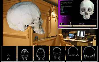 |
Skeletal specimens included in eSkeletons are digitized by a variety of methods at the High Resolution X-ray Computed Tomography Facility at the University of Texas at Austin (UTCT), a new partner in the NSF Tera Grid. Frame captures (at bottom) depict individual slices from the high resolution X-ray CT scan of the human skull.
Credit: John Kappelman, University of Texas at Austin |
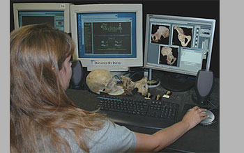 |
Production of the eSkeletons' web site depends upon the efforts of a large number of student research assistants. Kerri Wilhelm, a UT Austin undergraduate, reviews the anatomy of the human pelvis (os coxae) for correct labeling.
Credit: John Kappelman, University of Texas at Austin |
 |
John Kappelman also conducts paleontological field work at a variety of locations around the world. In the highlands of Ethiopia, he holds the fossilized teeth from a variety of extinct proboscideans (left hand) and the bizarre arsinothere (right hand). These fossils date to 27 millions years ago. See http://www.nsf.gov/od/lpa/news/03/pr03131.htm
Credit: Tab Rasmussen, Washington University, St. Louis |
|