|
Contents
First in a
Series:
Nutrition and Brain Function
Food for the Aging Mind
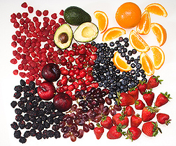
The oxygen radical absorbance capacity (ORAC) scores of fruits
vary. In this mix of fruit, the ORAC score of blueberries is highest, followed
by (in order) the scores of black plum, blackberries, raspberries,
strawberries, sweet cherries, avocado, navel orange, and red grapes.
(D833-1)
|
Scientists know that certain nutrients and other key chemical
compounds are essential to human brain function. Serious deficiencies in some
of these, such as vitamin B12 and iron, can lead to impaired cognitive function
due to neurological, or nerve fiber, complications.
Cognition can be defined as the ability to use
simple-to-complex information to meet the challenges of daily living.
So, could careful attention to diet help protect the aging
brain from problems with nerve cell signals involved in memory and cognition? A
clear-cut answer could greatly affect the 77 million baby boomers who are now
facing retirement. Their independence, quality of life, and even economic
status will largely be defined by their ability to traffic information signals
as they age.
In researching the nutrition-brain connection, new technologies
are being used, such as those that take images of the brain or actually count
individual brain cells. Behavioral tests that measure motor and cognitive
skills—or lack thereof—are also providing insights. Yet the science
of nutrition and brain function is relatively new and evolving.
Agricultural
Research Service scientists at several locations nationwide are
contributing to a growing body of research that explores the effect of diet and
nutrition on the brain and its function across the lifespan.

Biochemist Donna Bielinski prepares mammalian tissue samples to
look for the formation of new neurons, or neurogenesis.
(D835-1)
|
Boosting Neuronal Function
The brain’s billions of neurons “talk” to one
another through chemical neurotransmitters that convey signals through neural
pathways. These chemical transporters—which include norepinephrine,
serotonin, and dopamine—are key to signal movement.
Although people naturally lose brain cells throughout their
lives, the process of neuronal death does not necessarily accelerate with
aging. “There is a lot of individual difference,” says ARS
neuroscientist James Joseph. “Loss of mental agility may be less due to
loss of brain cells than to the cells’ failure to communicate
effectively.”
Joseph heads the Neuroscience Laboratory at the Jean Mayer USDA
Human Nutrition Research Center on Aging (HNRCA) at Tufts University in Boston.
There, researchers are looking at the beneficial effects of certain dietary
plant compounds to learn how they affect brain function.
“Vitamins and minerals in plant foods provide protective
antioxidants,” says Joseph. “But fruits, vegetables, nuts, seeds,
and grains contain thousands of other types of compounds that contribute
significantly to the overall dietary intake of antioxidants.
“A partial measure of the antioxidant effect is called
‘ORAC,’ for Oxygen Radical Absorbance Capacity. ORAC scores are now
showing up in charts and on some food and beverage packages. They may be
helpful in choosing foods to include in your diet.”
Perhaps there is no better place in which to gauge the power of
antioxidants than between the minute connections of the nerve cells.

ORAC (oxygen radical absorbance capacity) scores of vegetables
vary from plant to plant.
(D832-1)
|
Bucking Long-Held Dogma
Eight years ago, Joseph and colleagues began publishing a
series of studies, done in rodents, that shed light on the relationship between
various diets and the mechanisms behind cognitive losses in specific
neighborhoods of the aging brain.
Many in the series are groundbreaking in that they challenge
the long-accepted belief that the central nervous system, which includes the
brain, is not capable of regenerating itself. Other published studies in the
series echo similar findings based on primate and human brain research at the
Salk Institute for Biological Studies, San Diego, California. Scientists there,
using new technologies, disputed the notion that the brain does not make new
neurons—a process called “neurogenesis”—into old age:
It does, but at a much slower rate.
One of the first of Joseph’s studies, published in the
Journal of Neuroscience, showed a protective effect of
consuming antioxidants. Study rats were fed—from adulthood to middle
age—vitamin E, strawberry extracts, or spinach extracts, all with similar
ORAC values. Animals receiving the high-antioxidant diets did not experience
the age-related cognitive performance losses seen in control rats fed standard
chow.

Fruits being freeze-dried by technician John McEwen for use in
experimental diets.
(K8354-1)
|
A later study, also published in the Journal of
Neuroscience, showed a reversal of functional loss among rats on
special diets. Each of three groups of rats, equivalent in age to 63-year-old
humans, was fed a different high-antioxidant extract. A control group was fed
standard chow. After 8 weeks—equivalent to about 10 years in
humans—the rats’ performance levels were measured.
The rats fed the spinach, strawberry, or blueberry extracts
effectively reversed age-related deficits in neuronal and cognitive function.
In addition, the blueberry-fed group far outperformed their peers while
traversing a rotating rod to test balance and coordination.
“Despite their status as ‘senior citizens,’
those rats showed remarkable stamina on neuromotor function tests,” says
psychologist and coauthor Barbara Shukitt-Hale, also with the Neuroscience
Laboratory.
Examination of the brain tissue of those blueberry-fed rats
showed much higher levels of dopamine than were found in the other groups.
Dopamine has many functions within the brain. In particular, it can affect the
way the brain controls movements.
“We suspected that the combined antioxidant potency of
compounds in blueberry extract may have reduced inflammatory compounds in the
brains of these older animals,” says Joseph. “Inflammation
ordinarily contributes to neuronal and behavioral shortfalls during
aging.”
Tests have since shown that blueberry compounds cross the
blood-brain barrier and localize in rodent brain tissue.

Biologist Derek Fisher (left) and physiologist Jim Joseph
observe primary hippocampal neurons using fluorescent microscopy and real-time
calcium imaging to determine the effects of blueberry polyphenol treatments in
protecting against oxidative and inflammatory stress.
(D834-1)
|
Hard News: Brain Plaques
Later, the lab’s researchers published an
Alzheimer’s disease model study in Nutritional Neuroscience.
They studied mice that carried a genetic mutation for promoting increased
amounts of amyloid beta, a protein fragment found within the telltale neuritic
plaque, or “hardening of the brain,” seen in Alzheimer’s
disease.
Although the exact cause of Alzheimer’s is not completely
understood, experts have recently identified one mechanism involving the
insufficient breakdown and recycling of amyloid protein in the brain. That
mechanism is both genetic and physiological. In those individuals, normally
harmless amyloid protein turns into fragments of amyloid beta, which build up
as plaque in the brain rather than being escorted into cellular recycling. That
action leads to cell death and weakened neuronal communication.
In the mouse study, beginning at age 4 months—early
adulthood—half the brain-plaqued group was fed a diet that included
blueberry extract for 8 months. The other half was fed standard rat chow and so
was a control group of mice that didn’t carry the amyloid-plaque
mutation.
At 12 months—early middle age—all groups were
tested for their performance in a maze.

Fruits and vegetables high in antioxidants play a role in brain
function. Here, a shopper selects berries and other fruit for their ORAC
(Oxygen Radical Absorbance Capacity) value.
(D847-1)
|
The brain-plaqued mice that were fed the blueberry extract
performed as well as the healthy control mice and performed much better than
their brain-plaqued peers fed standard chow.
A look at the plaqued brains of both the blueberry-fed and
chow-fed mice after death revealed no difference in the number of brain plaques
in either group. “Amyloid-beta-induced plaques are only one aspect of
Alzheimer’s disease,” says Joseph. “But the fact that we saw
a diet-induced behavioral difference, despite a similarity in plaque density in
both these animal groups, is significant.”
The team found increased activity of a family of enzymes called
“kinases” in the brains of the amyloid-plaqued mice that were fed
blueberry extract. Two kinases found in particular, ERK and PKC, are important
in mediating cognitive function, such as converting short-term memory to
long-term.
“These kinase molecules are involved in signaling
pathways for learning and memory,” says Joseph. “It could be that
the increased kinase activity within the plaque-ridden brains of the
blueberry-fed mice enhanced the signaling in certain receptors.”
Brain Cells Are Born
Another HNRCA rat study looked at the aged brain’s
ability to change physiologically—a condition scientists refer to as
“neuronal plasticity.” In addition to cell division and
differentiation, or “mission assignment,” brain tissue undergoes
many other changes throughout aging.
For example, a newborn sprouts billions of nerve cells while
soaking up information from the environment. But lower levels of synapse growth
continue in waves throughout the lifespan. Little-used synapses are eliminated,
while others are strengthened in a neuronal pruning process, of sorts.
Repair mechanisms involve neural immune cells, called
“microglia,” that seek to heal and protect injured brain tissue;
enzymes that regulate safe chemical levels; and genes that are expressed in
response to inflammation.
The neuronal-plasticity study investigated the physiological
link between nutrition and the memory-control hippocampal area of the aged
brain. That region, in the center of the brain, is essential for what’s
called “working” or “short-term” memory. It receives
and processes data, and then, if needed, passes it on for storage.
Neurogenesis also plays a role in the formation of new
memories. The capacity of the hippocampus to produce new neurons is thought to
be greatly diminished during aging. But this study suggested that old rats fed
blueberry extracts for a short time had increased neurogenesis in the dentate
gyrus area of their brain’s hippocampus. The dentate gyrus is one of the
few regions of the brain where neurogenesis occurs.
“We found changes in the proliferation of neurons in
blueberry-fed rats,” said Gemma Casadesus, formerly a graduate student
with the Neuroscience Laboratory and now with Case Western Reserve University.
In maze tests, blueberry-fed aged lab rats showed improvement in cognition over
chow-fed peers.
“There was an association between the proliferation of neuronal precursor
cells and better performance of spatial memory,” she says.
A Test of Learning and
Memory
|
Fed standard chow
|
Early trial
|
Later trial
|
 D836-1
|

D836-2
|
Fed blueberry extract
|
Early trial
|
Later trial
|

D836-3
|
 D836-4
|
To measure cognition in rats, ARS scientists
used video imaging of a pool to track the route rats took to find a submerged
platform (the end point of their swim). These diagrams show the top views of
the routes taken. In the trials, the rats fed the standard chow took longer to
find the platform and showed little or no progress in learning. In the same
water maze, rats fed the blueberry extract learned to locate the platform
faster.
Click the images for more information and to download high-resolution
images.
|
The researchers don’t yet know whether the cognitive
improvements seen in the aged blueberry-fed rats translate to humans.
“But it’s an important step in learning about the brain’s
ability to rescue itself from age-associated declines in physiological
function,” Casadesus says.
Can You Hear Me Now?
Neurons that can’t get their messages through signaling
pathways are like cell phones that can’t get their signals through to
other cell phones. Why does this happen?
As the brain matures, cell division becomes largely restricted
to specific regions of the brain, and brain cells tend to become more
vulnerable to two partners in crime: oxidative stress and inflammation.
In the body, free radicals—weakened atoms formed during
activities of daily living—are missing an
electron and want to bond with neighboring biomolecules to stabilize. The
problem is that unless neutralized, free radicals cause cellular damage known
as “oxidative stress.”
Cellular antioxidant defense systems counterbalance these rogue
molecules, but they’re not 100 percent effective—particularly as
the body and brain mature. And the brain is thought to be especially vulnerable
to oxidative stress.
“Weighing just 3 pounds, the brain accounts for only 2
percent of the body’s total mass, yet it uses up to half of the
body’s total oxygen consumed during mental activity,” says Joseph.
“Phytochemicals, together with essential nutrients in foods, provide a
health-benefits cocktail of sorts. It is feasible that continued research in
this area will point to dietary regimens that are effective in boosting
neuronal function.”
Inflammation is thought to be stoked by the overactivation of
microglia—the neural immune cells mentioned earlier.
Microglia are usually dormant, but they migrate to the site of
any brain injury. These sentries make up about 20 percent of the cell
population in certain regions of the brain.
While seeking to protect and repair tissue, microglia cells
produce and send out molecular stress signals, some by way of defensive
cytokines, as a bugle call to other cells. Those signals begin a cascade of
reactions, including the activation of genes that express proteins and other
stress chemicals to help clear away cellular debris.
Microglial activation by amyloid beta is thought to be a key
event in the progression of Alzheimer’s disease. “When microglia
are stuck in an always-on loop in response to plaque buildup in the brain, they
become problematic in and of themselves,” says Joseph.
This year, Francis Lau, a molecular biologist in the
Neuroscience Laboratory, published a study that investigated whether blueberry
extracts could have a preventive effect on inflammatory signals coming from
activated microglia cells.
Microglial activation is considered the hallmark of
inflammation in the central nervous system. For this study, Lau used a rodent
microglial cell line that has previously served as a model to study
plaque-induced microglial activation.
Lau exposed groups of those test cells to various levels of
blueberry extracts. He then challenged the cells with oxidative stress by
exposing them to a toxin—lipopolysaccharide—that triggers secretion
of inflammatory chemicals.
Neuroinflammation has been linked to the expression of genes
that spew two inflammatory enzymes, iNOS and COX-2, and two cytokines, IL-1b
and TNF-a.
Lau used real-time PCR (polymerase chain reaction) to find and
measure expression of genes that produce iNOS and COX-2 in the stress-induced
cell cultures. He found that the blueberry treatment significantly reduced that
expression.
The blueberry extract also markedly lessened secretion of the
two inflammatory cytokines. In fact, says Lau, “In cells exposed to the
highest blueberry extract concentration, the amount of TNF-a cytokine found was
next to nothing—essentially identical to that found in the control
cells.”
Looking to the Future
The food industry is now using a range of new and existing
product ingredients to gain entrance into the emerging brain-health market.
Some are producing food labels that list ORAC values—for example, for use
on containers of polyphenol-rich fruit juices and teas. So far, however, there
has been no review conducted by the U.S. Food and Drug Administration on health
benefits from eating berries.
Future studies at HNRCA will ideally include use of new
diagnostic tools as well as human clinical trials. Neuroimaging equipment, for
example, could be used to monitor the influence of various dietary factors on
development of plaque within the human brain. Such studies aim to find the best
dietary regimens to help adults preserve their mental capabilities while
aging.—By Rosalie Marion
Bliss, Agricultural Research Service Information Staff.
This research is part of Human Nutrition, an ARS national
program (#107) described on the World Wide Web at
www.nps.ars.usda.gov.
James A.
Joseph is with the USDA-ARS Human
Nutrition Research Center on Aging at Tufts University, 711 Washington St.,
Boston, MA 02111; phone (617) 556-3178, fax (617) 556-3222.
"Nutrition and Brain Function" was
published in the August 2007
issue of Agricultural Research magazine.
[Top]
|