|

All Images
Discovery
From Fingerprints to Fiberprints

Back to article | Note about images
 |
This thumbprint appeared after researchers sprayed it with a super glue, a process that forensic scientists use at crime scenes. The print is visible because chemicals in the residue from the finger reacted with the glue to form tiny, 3-dimensional structures.
Credit: Greg Greico, Penn State |
Download the high-resolution JPG version of the image. (1.4 MB)
|
Use your mouse to right-click (or Ctrl-click on a Mac) the link above and choose the option that will save the file or target to your computer.
|
 |
Polymer fibers grow on a fingerprint at 30 degrees Celsius and a relative humidity of more than 95 percent. Image (a) is a low-magnification view, and image (b) shows a close-up view of the same (the inset shows the top view of fiber).
Credit: Reproduced by permission of The Royal Society of Chemistry; Penn State |
Download the high-resolution JPG version of the image. (44 KB)
|
Use your mouse to right-click (or Ctrl-click on a Mac) the link above and choose the option that will save the file or target to your computer.
|
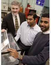 |
From left to right, the Pennsylvania State University team that studied the fingerprint-cyanoacrylate relationship: Henry C. Foley, professor of chemical engineering and director of the project; Pratik J. Mankidy, doctoral candidate in chemical engineering; and Ramakrishnan Rajagopalan, research associate.
Credit: Greg Greico, Penn State |
Download the high-resolution JPG version of the image. (1.2 MB)
|
Use your mouse to right-click (or Ctrl-click on a Mac) the link above and choose the option that will save the file or target to your computer.
|
 |
Henry C. Foley, professor of chemical engineering at Pennsylvania State University, directs the cyanoacrylate-fiber study.
Credit: Greg Greico, Penn State |
Download the high-resolution JPG version of the image. (1 MB)
|
Use your mouse to right-click (or Ctrl-click on a Mac) the link above and choose the option that will save the file or target to your computer.
|
 |
Researchers use sodium hydroxide to initiate the chemical reaction with the cyanoacrylate to create "tortellini-like" polymer films.
Credit: Reproduced by permission of The Royal Society of Chemistry; Penn State |
 |
Researchers grew these polymer nanofibers using a synthetic starting mixture of linoleic acid and salt water. The inset showing the same area at a higher magnification.
Credit: Reproduced by permission of The Royal Society of Chemistry; Penn State |
Download the high-resolution JPG version of the image. (46 KB)
|
Use your mouse to right-click (or Ctrl-click on a Mac) the link above and choose the option that will save the file or target to your computer.
|
 |
Nanofibers of poly (ethyl 2-cyanoacrylate) were grown on fingerprint ridges at 30 degrees Celsius at a relative humidity of more than 95 percent over a period of 16 hours. Scanning electron microscope image (a) shows a low magnification view; image (b) shows a close-up view of the ridge pattern; image (c) shows a close-up view of the nanofibers; and image (d) is a magnified view of a single fiber.
Credit: Reproduced by permission of The Royal Society of Chemistry; Penn State |
Download the high-resolution JPG version of the image. (128 KB)
|
Use your mouse to right-click (or Ctrl-click on a Mac) the link above and choose the option that will save the file or target to your computer.
|
|