What Is a Cataract?

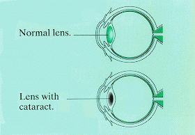
A cataract is a cloudy area in the lens of the eye that affects vision.
A normal lens is clear. It lets light pass to the retina, the light-sensitive tissue at the back of the eye. As a cataract develops, it becomes harder for a person to see. Vision may become cloudy or blurry, and colors may fade.
Most people have a cataract in both eyes. One eye may be worse than the other, however, because each cataract develops at a different rate. Some people with a cataract don't even know it. Their cataract may be small, or the changes in their vision may not bother them very much. Other people cannot see well enough to do the things they need or want to do.
Who Is Affected?
Most cataracts are related to aging. About half of Americans ages 65 to 74 have a cataract. About 70 percent of those age 75 and older have this condition. However, other conditions can increase the risk of developing cataracts. Examples include diabetes, smoking and alcohol use, and prolonged exposure to sunlight.
How Is a Cataract Treated?
Surgery is used to remove a cataract when vision loss interferes with everyday activities such as driving, reading, or watching TV. If you have cataracts in both eyes, the surgery will be performed on each eye at separate times, usually 4 to 8 weeks apart. In 90 percent of cases, people who have cataract surgery have better vision afterwards.
Proceed to Next Section

