 |
|
|
||||
| Honolulu Field Station | ||||
 |
Background: Coral reefs worldwide are under tremendous stress primarily due to human activities along the coasts. While climate change, overfishing and coastal development have been implicated as a major cause of coral reef decline, diseases seem to play an increasing role. A prime example of this is the Western Atlantic where various diseases have killed hard and soft corals throughout the Carribean. Unfortunately, only a few of these diseases have been described in any meaningful detail. In part, this is because not much is known about the physiology or biology of many coral species. |
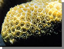 |
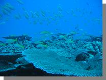
Corals are animals: When you are snorkeling or diving over a coral reef, almost all of what you see is alive. You probably notice the large things (fish, urchins) but most of what is on the reef are things like algae and corals. Algae are plants, but corals are not. Corals are actually related to jellyfish and are known as colonial organisms (many individuals making up a single large organism). If you look closely at a coral colony, you will see numerous cup-shaped structure surrounded by little arms. Each of these is known as a polyp and consists of a mouth (cup like structure) surrounded by tentacles (little arms). Depending on the species, a coral colony can have dozens to thousands of these polyps, all interconnected, and all feeding on microscopic material in the water column. One other thing that makes corals unique is that they have little algal cells in their tissues (zooxanthella) that provide essential nutrients to the polyp. In return, the polyp provides a safe habitat from the algae. The polyps and surrounding tissues secrete the skeleton which forms the major structure of coral reefs. So, next time you're cruising over a reef, recognize that you're looking at a thin layer of living tissue covering inert skeleton. This is why people are encouraged not to touch or stand on reefs, because wherever you touch, you risk damaging fragile tissue. |
 |
Corals and disease and confusion: Because corals are animals, they are susceptible to all the things that can cause illness or death in other groups of animals. Diseases in corals can be due to both transmissible agents (microbes) and non-infectious things like toxicants. Disease has played the most visible role in corals from the Western Atlantic. There, loss of major reef building species of corals has been attributed to syndromes like white band, black band and white blotch. Unfortunately, many diseases in corals are described based on descriptions of clinical signs seen in the field. Relatively few diseases in corals have been described completely (both from the morphologic standpoint and with an eye to determining what is causing clinical signs (lesions) in corals. Hence, the scientific literature on diseases of corals is full of different names for syndromes that could be caused by different or the same factors. |
 |
A collaborative project : The Honolulu Field Station has been looking at one facet of coral disease, namely, devising ways to systematically describe the morphology of lesions seen in corals. A good description of morphologic changes at the gross and cellular level provide the foundation for any description of any disease, and this seems to be lacking in the coral disease literature. Limited surveys of coral diseases have been done on Oahu and French Frigate Shoals and Johnston Atoll in the Central Pacific. These efforts, however, were limited to examining pathology of corals, however, many other facets of disease investigation in corals were overlooked due to lack of resources. This year, the Hawaii Coral Reef Initiative awarded funding to a collaborative team composed of the Hawaii Institute of Marine Biology, Hawaii Division of Aquatic Resources, Bishop Museum and the Honolulu Field Station to take a more comprehensive look at the status of coral health around Oahu by incorporating field surveys with laboratory methods such as microscopic examinations and molecular analyses. |
|
Nomenclature: We have argued that in most cases, it is very difficult to know what is causing a lesion on a coral by simple field observation. Thus, we have simplified our nomenclature to describing morphologic changes that occur in the corals. For the purposes of classifying lesions in the field, we categorize lesions in corals as: |
 |
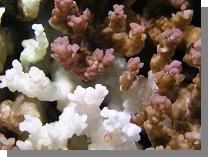
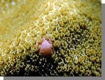
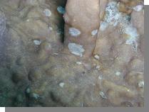
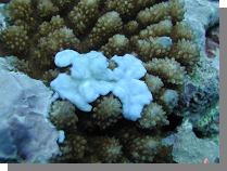
Discoloration: This is where there is a change in color from normal in the coral tissue. While this often applies to what is known as bleaching (loss of pigmentation in the tissues revealing the white skeleton underneath), it can apply to any coral manifesting areas of tissue discoloration (palor, complete loss of pigment, or change in pigmentation). |
 |
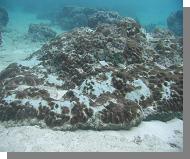
Tissue loss: This is where there is absence of tissue revealing bare or algae covered skeleton. |
 |
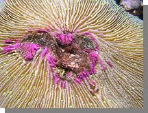
Growth anomalies : This is where there is abnormal growth of skeleton or tissue. |
|
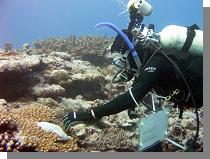
Quantifying lesions: Lesions are quantified on a reef using a combination of photoquadrats and 25 m line transects. Photos are taken along a 25 m transect and these are used to quantify the substrate and percent cover of each type of coral (down to genus). Coral colonies are also counted on a 25 m X 1 m belt and lesions classified as above. Finally, lesions only are counted on a 6 X 25 m belt along the transect. These bits of information are then used to assess the percentage of different genera of corals affected by different lesion types. |
|
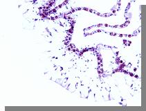
Describing lesions: Lesions are photographed using pan and close-up digital photography. Samples of lesions are taken in a fixative solution, the skeleton dissolved using acid solution, and the tissues placed on microscope slides. Cellular changes associated with the lesions are then described. This process (histopathology) can sometimes reveal things like parasites, bacteria, or micro-algae that interact with coral tissue to cause death. Lesions are classified based on microscopic observations and these microscopic descriptions are related back to what was seen grossly. |
| AccessibilityFOIAPrivacyPolicies and Notices | |
 | |